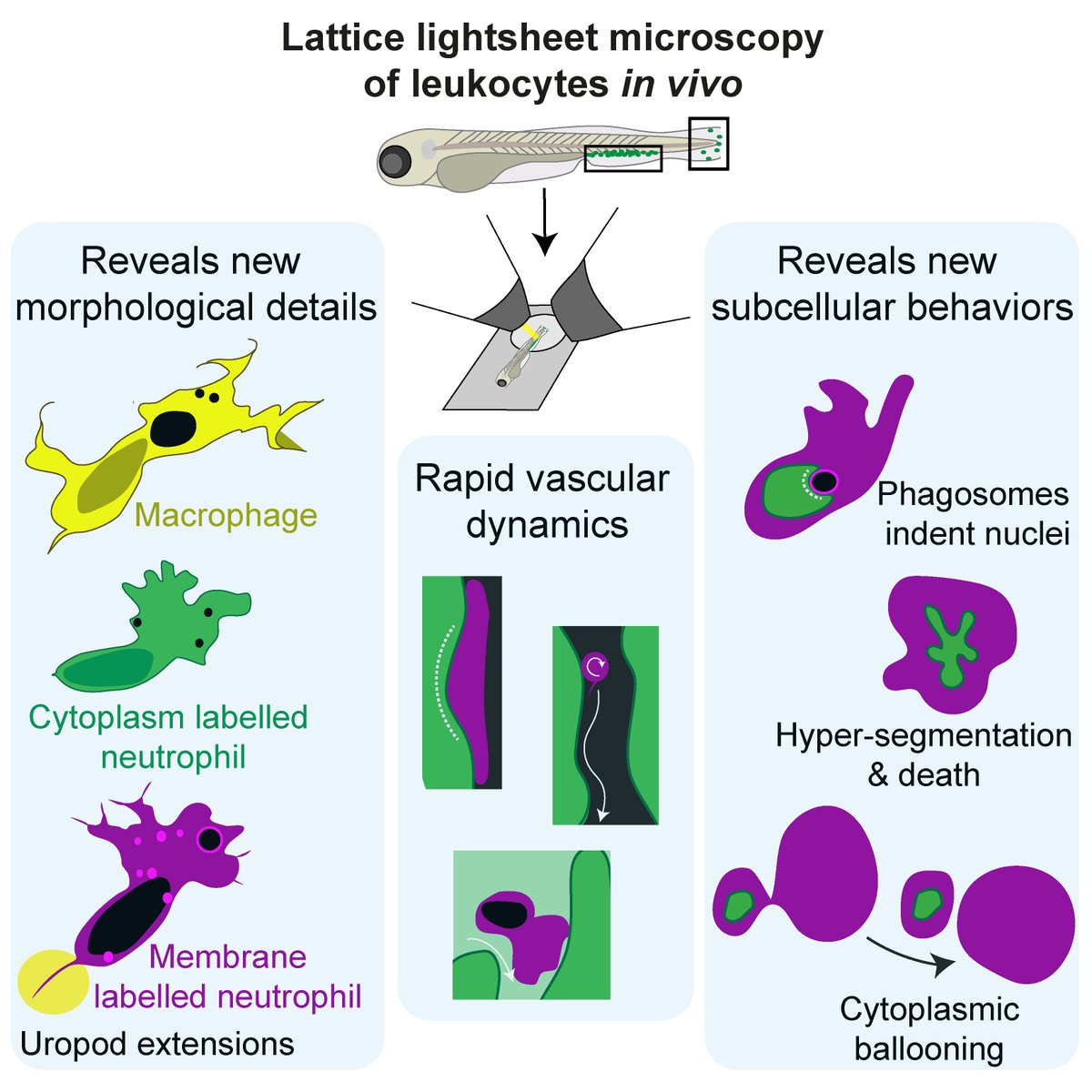
Our new paper jam-packed with exciting lattice #lightsheet microscopy is online! Imaging #zebrafish leukocytes, we reveal new dynamic cellular & subcellular features in vivo. At this resolution, these fast‐moving cells are captured in all their glory!/1 https://doi.org/10.1002/JLB.3HI0120-589R">https://doi.org/10.1002/J...
Focusing on #neutrophil & #macrophage migration, we characterized cell morphology beyond in vivo confocal blobs & blurs: using 4-17s/stack, 0.263µm slices!
Membrane-labelling is clearly superior as it highlights thin cytoplasmic extensions, and internal vacuoles and vesicles /2
Membrane-labelling is clearly superior as it highlights thin cytoplasmic extensions, and internal vacuoles and vesicles /2
As #neutrophils are especially fast and difficult to image in vivo, we refined their morphological description by imaging their ~200nm-thin trailing extensions. These prevalent uropod extensions can reach ~25um in length, leave deposits behind, and are mediated by myosin II /3
Intra- and extra-vascular neutrophils (magenta) in blood vessel environments (green) displayed blood-flow-induced migration modes and collisions, and unexpected leukocyte-induced endothelial deformation. We also captured classic rolling neutrophils with cytoplasmic tethers /4
Subcellular events captured with membrane-labelled neutrophils (magenta) & a nuclear envelope reporter (green) showed nuclei indent phagosomes, the nuclei of dying cells hypersegment. Lastly, novel cytoplasmic ballooning shows how this imaging can reveal weird new behaviors! https://abs.twimg.com/emoji/v2/... draggable="false" alt="😀" title="Grinning face" aria-label="Emoji: Grinning face"> /5
Huge thanks to all my coauthors @KeightleyCris @JawnnyH @ScopeShifu @ARMI_Labs , and to all the crew at @AICjanelia for my visit which truly propelled this work forward! And if you read this thread and check out the paper, thank you!  https://abs.twimg.com/emoji/v2/... draggable="false" alt="🔬" title="Microscope" aria-label="Emoji: Microscope">
 https://abs.twimg.com/emoji/v2/... draggable="false" alt="🐟" title="Fish" aria-label="Emoji: Fish">
 https://abs.twimg.com/emoji/v2/... draggable="false" alt="👩🔬" title="Woman scientist" aria-label="Emoji: Woman scientist"> /6

 Read on Twitter


