Thread:
The 10 publications below provide scientific evidence that patients with #MECFS have #brain abnormalities in #neuroinflammation, #metabolism, #neurological connections and blood perfusion
#pwME suffer from #chronicillness
#pwME are #SickNotWeak
The 10 publications below provide scientific evidence that patients with #MECFS have #brain abnormalities in #neuroinflammation, #metabolism, #neurological connections and blood perfusion
#pwME suffer from #chronicillness
#pwME are #SickNotWeak
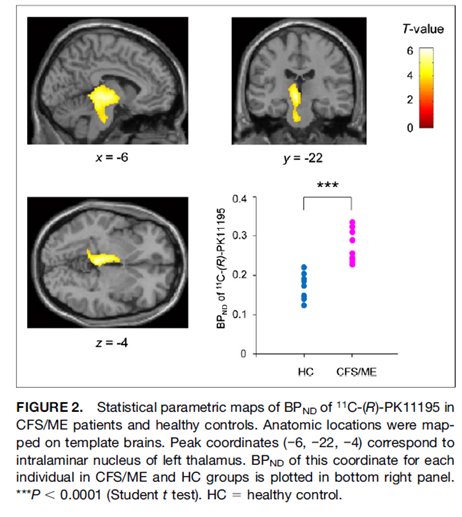
1. These studies “provide evidence of #neuroinflammation in #MECFS.. as well as evidence of the possible contribution of neuroinflammation to the pathophysiology of #MECFS”
#pwME
http://jnm.snmjournals.org/content/55/6/945.long">https://jnm.snmjournals.org/content/5...
#pwME
http://jnm.snmjournals.org/content/55/6/945.long">https://jnm.snmjournals.org/content/5...
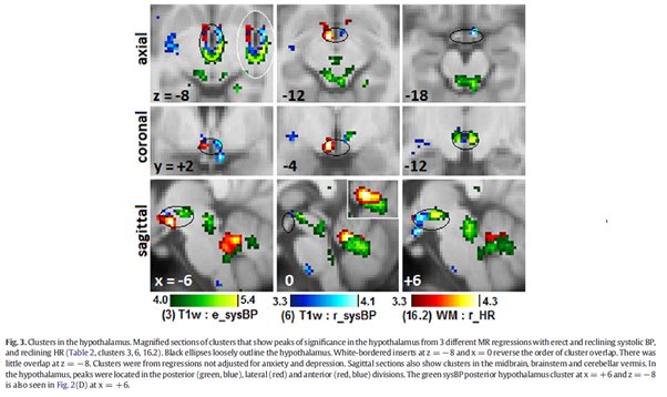
2. This Australian study found abnormalities in the #brain MRIs and peripheral Blood Pressure and Heart Rate in #MECFS patients including the Vasomotor centre, midbrain and hypothalamus
#pwME
https://www.sciencedirect.com/science/article/pii/S2213158216300584">https://www.sciencedirect.com/science/a...
#pwME
https://www.sciencedirect.com/science/article/pii/S2213158216300584">https://www.sciencedirect.com/science/a...
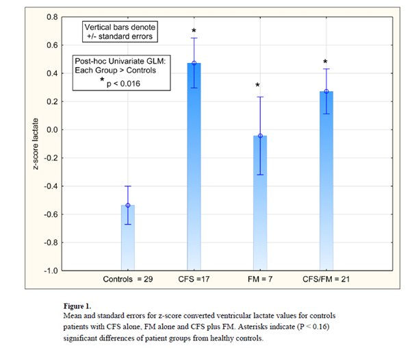
3. Brain MRIs show that patients with #MECFS have significantly higher lactate in their cerebral spinal fluid compared to healthy controls
These findings suggest that #pwME have a problem in brain-related mitochondrial #metabolism
https://www.ncbi.nlm.nih.gov/pubmed/29308330 ">https://www.ncbi.nlm.nih.gov/pubmed/29...
These findings suggest that #pwME have a problem in brain-related mitochondrial #metabolism
https://www.ncbi.nlm.nih.gov/pubmed/29308330 ">https://www.ncbi.nlm.nih.gov/pubmed/29...
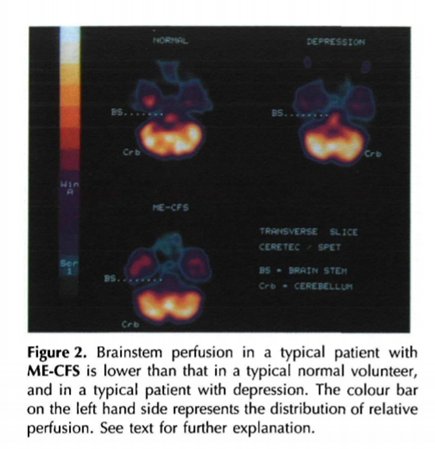
4. SPET analysis of #CFS patients found a reduction in brain perfusion
“our data suggest that brainstem hypoperfusion in #CFS patients could be due to an organic abnormality”
#MECFS #pwME
https://academic.oup.com/qjmed/article-abstract/88/11/767/1569403#.XEkeFLn1j5U.twitter">https://academic.oup.com/qjmed/art...
“our data suggest that brainstem hypoperfusion in #CFS patients could be due to an organic abnormality”
#MECFS #pwME
https://academic.oup.com/qjmed/article-abstract/88/11/767/1569403#.XEkeFLn1j5U.twitter">https://academic.oup.com/qjmed/art...
5. These MRI studies found that the “impaired brainstem signal conduction [in #CFS patients] may stimulate elevated sensorimotor myelination.. [that may] have broad consequences for regulation of cerebral function..”
#MECFS #pwME
https://www.sciencedirect.com/science/article/pii/S2213158218302237">https://www.sciencedirect.com/science/a...
#MECFS #pwME
https://www.sciencedirect.com/science/article/pii/S2213158218302237">https://www.sciencedirect.com/science/a...
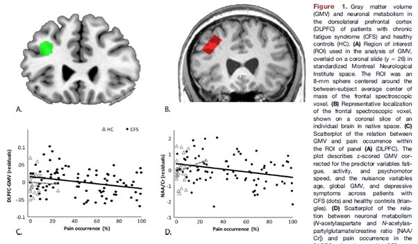
6. #Brain MRI studies in women with #CFS showed that the presence of pain is associated with neuronal alterations and #metabolic defects in the dorsolateral prefrontal cortex
#MECFS #pwME
https://www.biologicalpsychiatryjournal.com/article/S0006-3223(16)32737-8/abstract">https://www.biologicalpsychiatryjournal.com/article/S...
#MECFS #pwME
https://www.biologicalpsychiatryjournal.com/article/S0006-3223(16)32737-8/abstract">https://www.biologicalpsychiatryjournal.com/article/S...
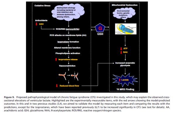
7. This MRI study found elevated lactate and reduced cortical glutathione (GSH) in the brains of #CFS patients suggesting increased oxidative stress, cerebral hypoperfusion and/or secondary #mitochondria dysfunction
#MECFS #pwME
https://onlinelibrary.wiley.com/doi/abs/10.1002/nbm.2772#.XEk4C8bECGg.twitter">https://onlinelibrary.wiley.com/doi/abs/1...
#MECFS #pwME
https://onlinelibrary.wiley.com/doi/abs/10.1002/nbm.2772#.XEk4C8bECGg.twitter">https://onlinelibrary.wiley.com/doi/abs/1...
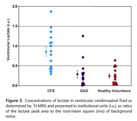
8. #Brain proton MRS imaging has shown that patients with #CFS had significantly increased ventricular lactate compared to healthy controls or patients with generalized anxiety disorder (GAD)
#MECFS #pwME
https://onlinelibrary.wiley.com/doi/abs/10.1002/nbm.2772#.XEk75i1YJ9Y.twitter">https://onlinelibrary.wiley.com/doi/abs/1...
#MECFS #pwME
https://onlinelibrary.wiley.com/doi/abs/10.1002/nbm.2772#.XEk75i1YJ9Y.twitter">https://onlinelibrary.wiley.com/doi/abs/1...
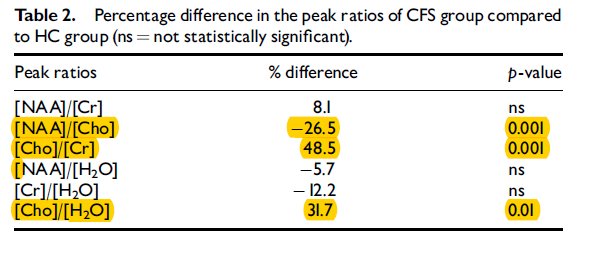
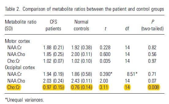
9. proton MRS analysis of the basal ganglia of #CFS brains found abnormal levels of N-acetyl aspartate, creatinine and choline-containing compounds
#MECFS #pwME
https://insights.ovid.com/pubmed?pmid=12598734">https://insights.ovid.com/pubmed...
#MECFS #pwME
https://insights.ovid.com/pubmed?pmid=12598734">https://insights.ovid.com/pubmed...
10. #Brain proton magnetic resonance spectroscopy found biochemical abnormalities in phospholipid #metabolism of #CFS brains
#MECFS #pwME
…https://onlinelibrary-wiley-com.ezproxy.lib.monash.edu.au/doi/epdf/10.1034/j.1600-0447.2002.01300.x">https://onlinelibrary-wiley-com.ezproxy.lib.monash.edu.au/doi/epdf/...
#MECFS #pwME
…https://onlinelibrary-wiley-com.ezproxy.lib.monash.edu.au/doi/epdf/10.1034/j.1600-0447.2002.01300.x">https://onlinelibrary-wiley-com.ezproxy.lib.monash.edu.au/doi/epdf/...
11. Conclusion: The above 10 research publications indicate that there are clear biochemical, metabolic and inflammatory abnormalities in the brains of #pwME
12. This @FrontNeurol study provides important guidelines and methodological approaches for future brain imaging studies examining #neuroinflammation in #MECFS https://www.frontiersin.org/article/10.3389/fneur.2018.01033">https://www.frontiersin.org/article/1...

 Read on Twitter
![5. These MRI studies found that the “impaired brainstem signal conduction [in #CFS patients] may stimulate elevated sensorimotor myelination.. [that may] have broad consequences for regulation of cerebral function..” #MECFS #pwME https://www.sciencedirect.com/science/a... 5. These MRI studies found that the “impaired brainstem signal conduction [in #CFS patients] may stimulate elevated sensorimotor myelination.. [that may] have broad consequences for regulation of cerebral function..” #MECFS #pwME https://www.sciencedirect.com/science/a...](https://pbs.twimg.com/media/Dx0JG44VAAcu9b5.jpg)
