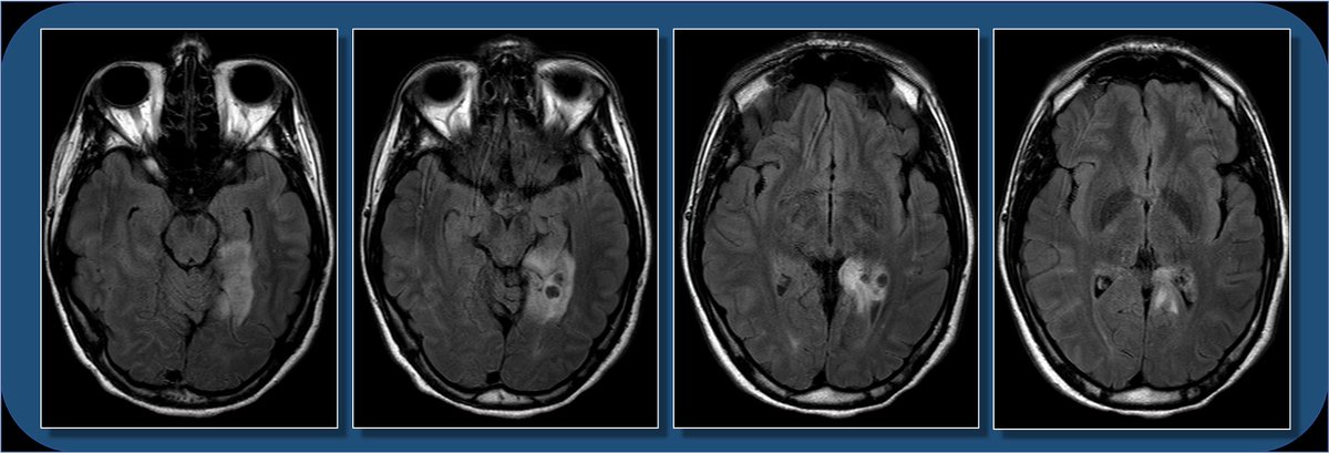
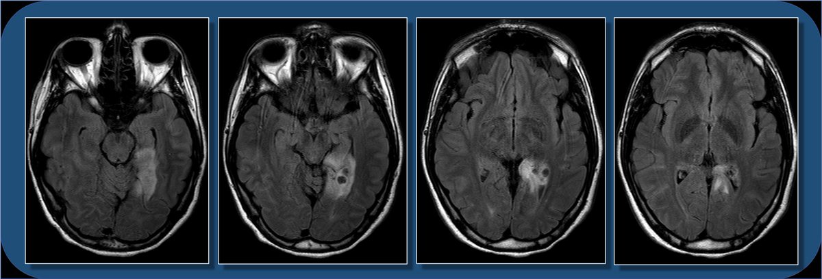
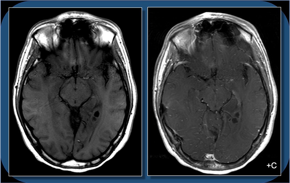
Hey #radres and #MEDstudents - a #neurorad case for you today. 18yoF w/ seizure. What do you see? What is the ddx? And WHERE is the lesion? Case explanation and gyral #neuroanatomy explanation coming soon.
#meded #medtwitter #FOAMed #FOAMrad #radiology #neurology #neurosurgery
#meded #medtwitter #FOAMed #FOAMrad #radiology #neurology #neurosurgery
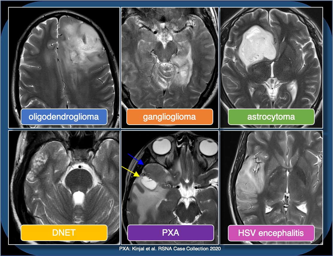
What is the LEAST likely diagnosis?
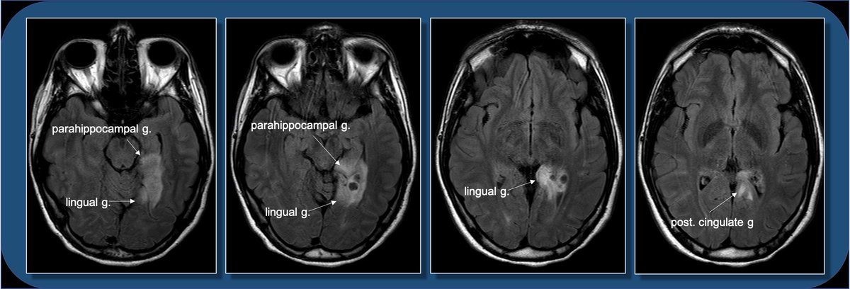
Which is not involved by the lesion?
18yoF with seizures. The FLAIR images show focal cortical expansion with cystic components, suggesting a cortically based tumor.
1/13
#radres #MEDstudents #neurorad #neuroanatomy #meded #medtwitter #FOAMed #FOAMrad #radiology #neurology #neurosurgery
1/13
#radres #MEDstudents #neurorad #neuroanatomy #meded #medtwitter #FOAMed #FOAMrad #radiology #neurology #neurosurgery
T1 images show mild T1 hypointense mass with cystic components, and minimal if any enhancement. So what’s the differential for a cortically based tumor in a child or young adult who presents with seizure? Pathology following resection c/w ganglioglioma.
2/13
2/13
Ddx for cortically based tumors in child/young adult includes oligodendroglioma (usually middle-aged adults, often with Ca++), DNET (bubbly, uncommon enhancement), PXA (prominent nodular enhancement), and astrocytoma (usually low grade). Encephalitis can also look similar.
3/13
3/13
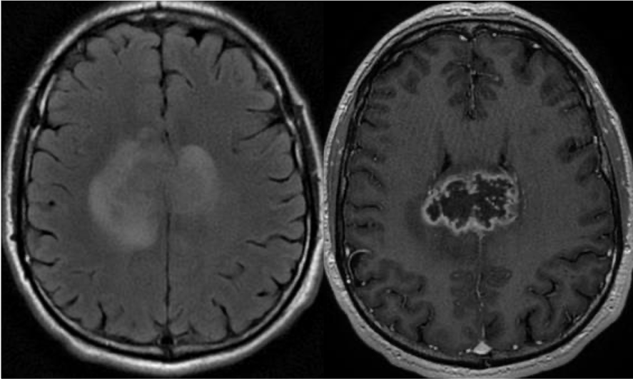
This is less likely to be a glioblastoma, which is usually centered in supratentorial WM, with thick peripheral enhancement, and centrally necrotic.
4/13
4/13
This location can be a bit difficult to describe if you aren’t up on gyral anatomy. Is it med temporal lobe? occipital lobe? limbic? Describing gyral location is more precise, can explain expected symptoms, and clue in future surgical approach.
5/13
5/13
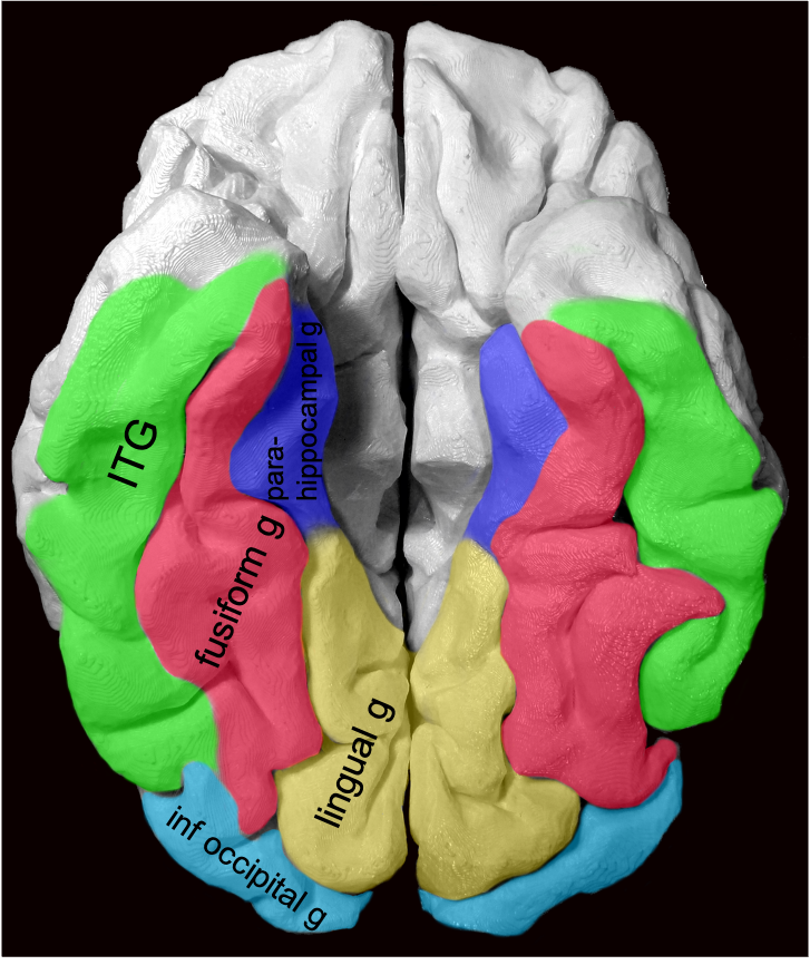
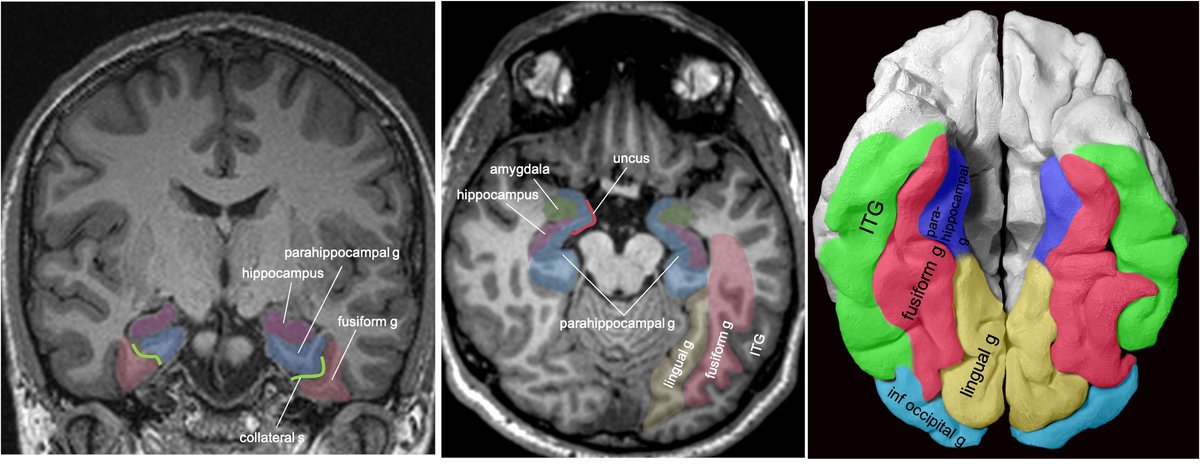
Gyral anatomy of inf occipitotemporal surface: The inf. temp. gyrus wraps from lateral https://abs.twimg.com/emoji/v2/... draggable="false" alt="➡️" title="Pfeil nach rechts" aria-label="Emoji: Pfeil nach rechts">inferior. The fusiform and lingual gyri span from occipital lobe posteriorly to temporal lobe anteriorly. Parahippocampal is medial temporal (anterior continuation of the lingual g).
6/13
6/13
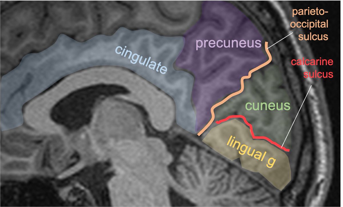
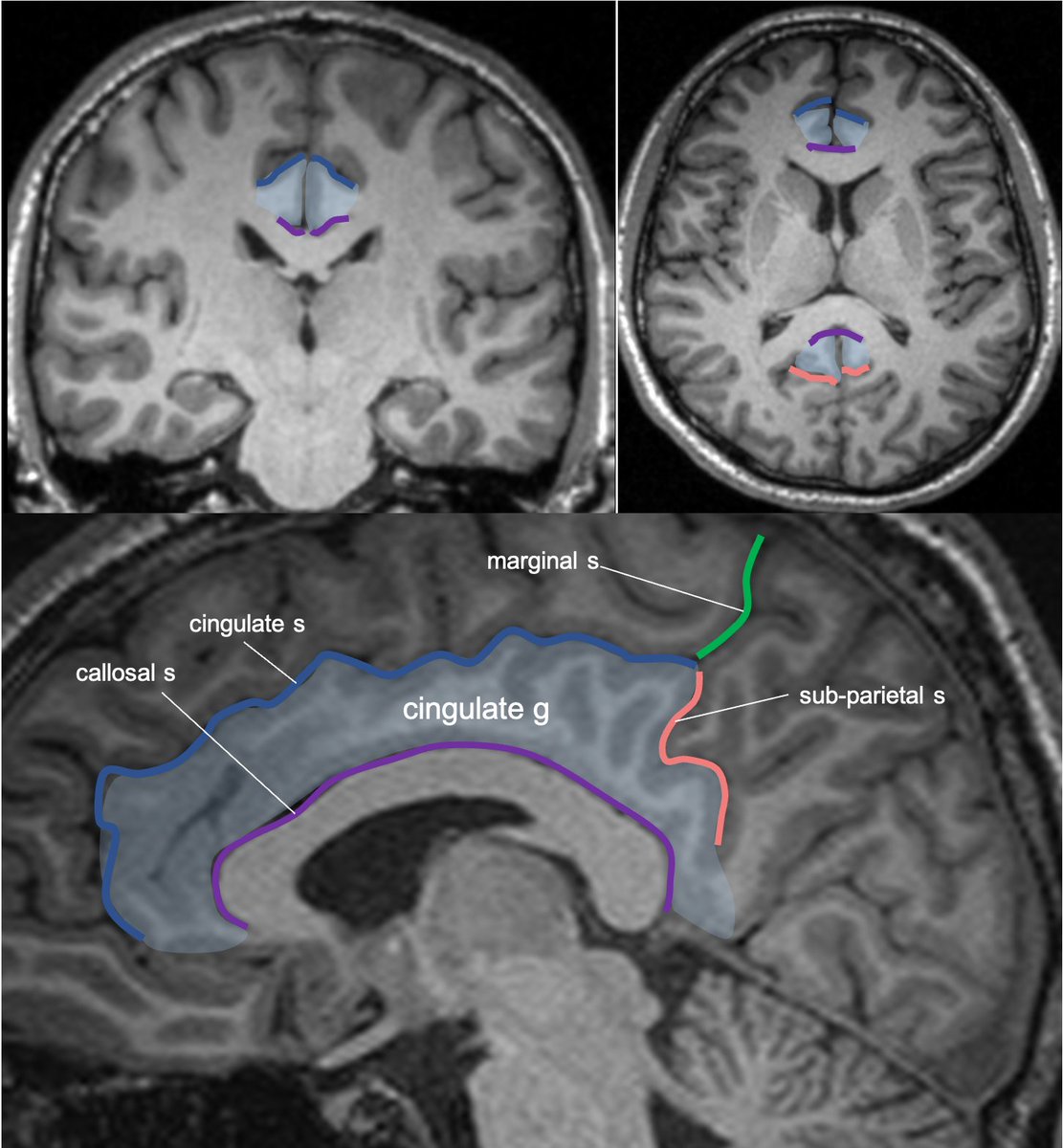
Gyral anatomy of the medial brain surface: calcarine sulcus divides the cuneus from lingual gyrus at the med occ lobe. The lingual g continues anteriorly along the med/ inferior temporal lobe. The cingulate g wraps sup & posterior to corpus callosum along the medial brain.
7/13
7/13
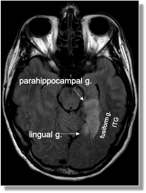
The lingual gyrus is involved in holistic visual and word processing, encoding visual memories, imagery, and dreaming. The ganglioglioma in the shown case is centered in the lingual gyrus.
8/13
8/13
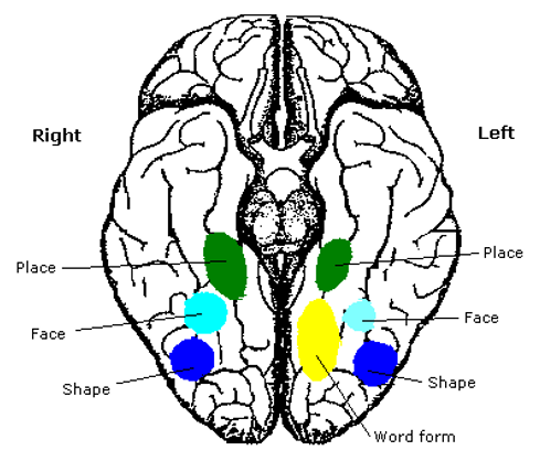
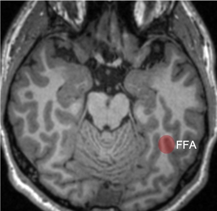
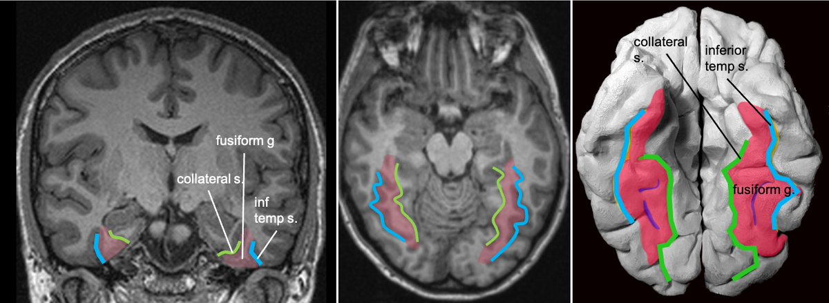
Fusiform g is just lateral to lingual g on the inf surface of temporal lobe, separated by collateral sulcus. It& #39;s medial to the ITG, separated by ITS. Involved in face and body recognition; word form recognition (on left). (Fusiform g was not definitively involved by tumor). 9/13
Parahippocampal g. is anterior continuation of lingual g. Part of Limbic System. Important in memory encoding and retrieval, visual and social contextualizing.
10/13
10/13
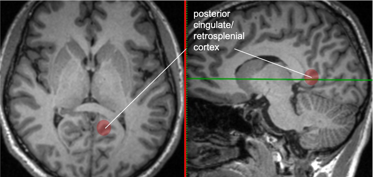
Cingulate g. is part of the limbic circuit; the post cingulate cortex (PCC) is important in default mode network, awareness, pain, and episodic memory retrieval. PCC including the retrosplenial cortex (which together some call restrosplenial gyrus) is involved by the tumor. 11/13
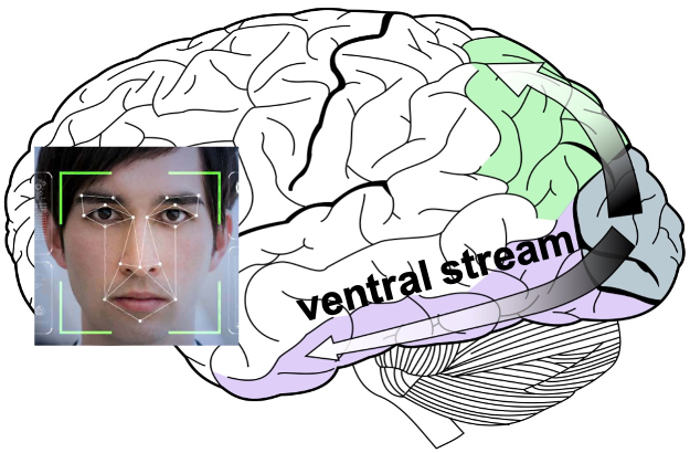
Some functional #neuroscience: Inf temp lobe is involved in higher order visual processing along the ventral stream (the "what is it" path of visual sensory processing https://abs.twimg.com/emoji/v2/... draggable="false" alt="➡️" title="Pfeil nach rechts" aria-label="Emoji: Pfeil nach rechts"> color/object/word/face/body perception/recognition/identification, & associated cognitive functions)
12/13
12/13

 Read on Twitter
 inferior. The fusiform and lingual gyri span from occipital lobe posteriorly to temporal lobe anteriorly. Parahippocampal is medial temporal (anterior continuation of the lingual g).6/13" title="Gyral anatomy of inf occipitotemporal surface: The inf. temp. gyrus wraps from lateralhttps://abs.twimg.com/emoji/v2/... draggable="false" alt="➡️" title="Pfeil nach rechts" aria-label="Emoji: Pfeil nach rechts">inferior. The fusiform and lingual gyri span from occipital lobe posteriorly to temporal lobe anteriorly. Parahippocampal is medial temporal (anterior continuation of the lingual g).6/13" class="img-responsive" style="max-width:100%;"/>


 color/object/word/face/body perception/recognition/identification, & associated cognitive functions)12/13" title="Some functional #neuroscience: Inf temp lobe is involved in higher order visual processing along the ventral stream (the "what is it" path of visual sensory processinghttps://abs.twimg.com/emoji/v2/... draggable="false" alt="➡️" title="Pfeil nach rechts" aria-label="Emoji: Pfeil nach rechts"> color/object/word/face/body perception/recognition/identification, & associated cognitive functions)12/13" class="img-responsive" style="max-width:100%;"/>
