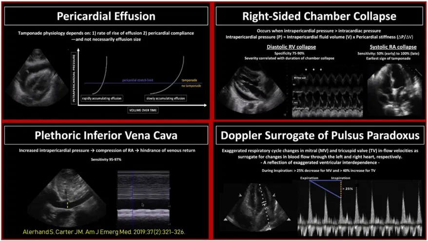
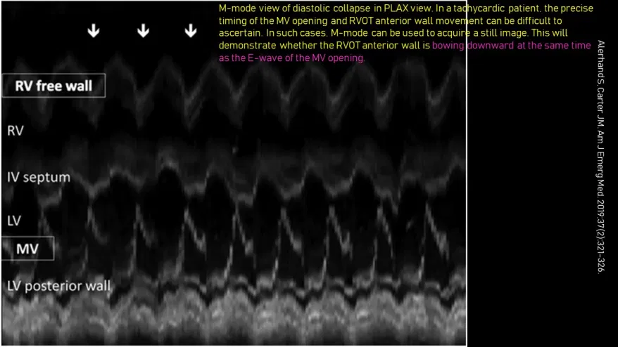
#POCUS #echofirst image of the day: #Nephrology consulted for a patient on #dialysis who presents with generalized fluid overload after missing a few sessions.
#physicalexam with sonoscope reveals this https://abs.twimg.com/emoji/v2/... draggable="false" alt="👇" title="Rückhand Zeigefinger nach unten" aria-label="Emoji: Rückhand Zeigefinger nach unten"> (much better than stethophone/wheeze detector
  https://abs.twimg.com/emoji/v2/... draggable="false" alt="😬" title="Grimasse schneidendes Gesicht" aria-label="Emoji: Grimasse schneidendes Gesicht">)
More images in thread https://abs.twimg.com/emoji/v2/... draggable="false" alt="🧵" title="Thread" aria-label="Emoji: Thread">
#physicalexam with sonoscope reveals this
More images in thread
#POCUS not only diagnoses effusion, but also helps to get an idea of severity, assess chamber collapse/tamponade physiology, PLUS facilitates #patienteducation. This pt also had LVH, mitral annular calcification (arrowhead) - now better understands importance of BP/ phos control.
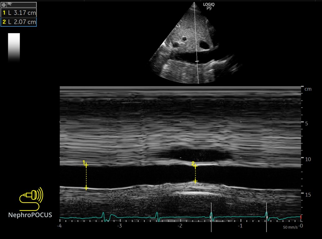
Big IVC with a little respiratory variation  https://abs.twimg.com/emoji/v2/... draggable="false" alt="👇" title="Rückhand Zeigefinger nach unten" aria-label="Emoji: Rückhand Zeigefinger nach unten"> #POCUS
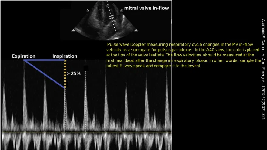
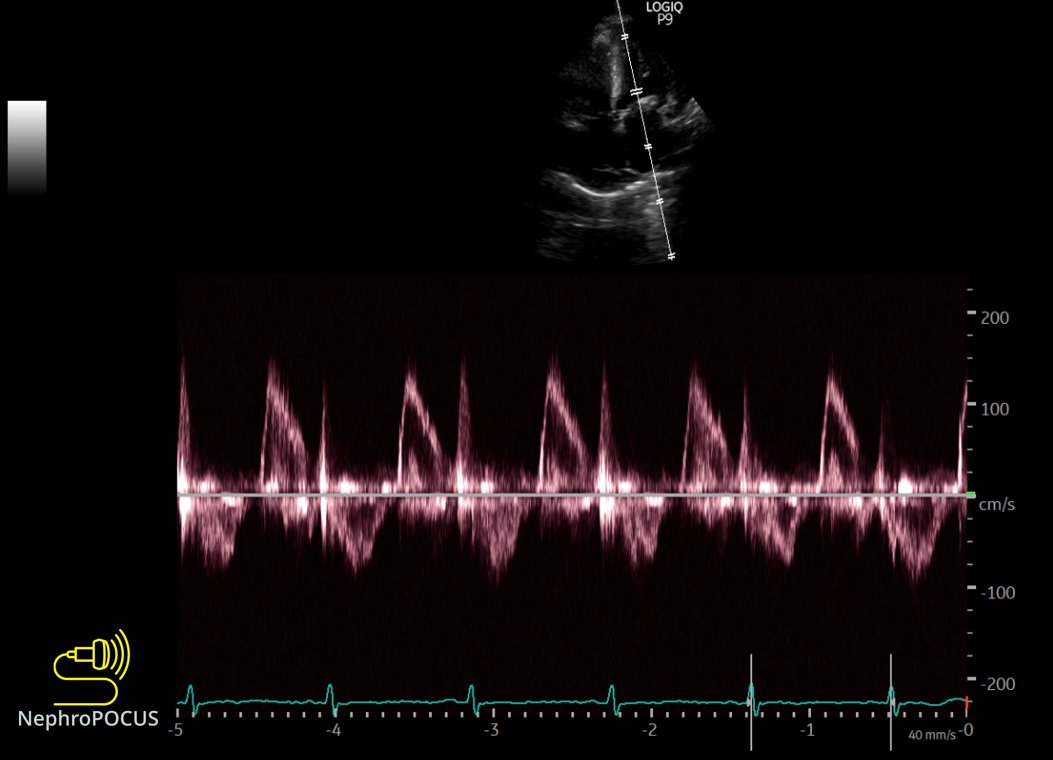
Mitral inflow Doppler #echofirst to assess echocardiographic pulsus paradoxus  https://abs.twimg.com/emoji/v2/... draggable="false" alt="👇" title="Rückhand Zeigefinger nach unten" aria-label="Emoji: Rückhand Zeigefinger nach unten">
Obtained during quiet breathing; no sniff
Obtained during quiet breathing; no sniff
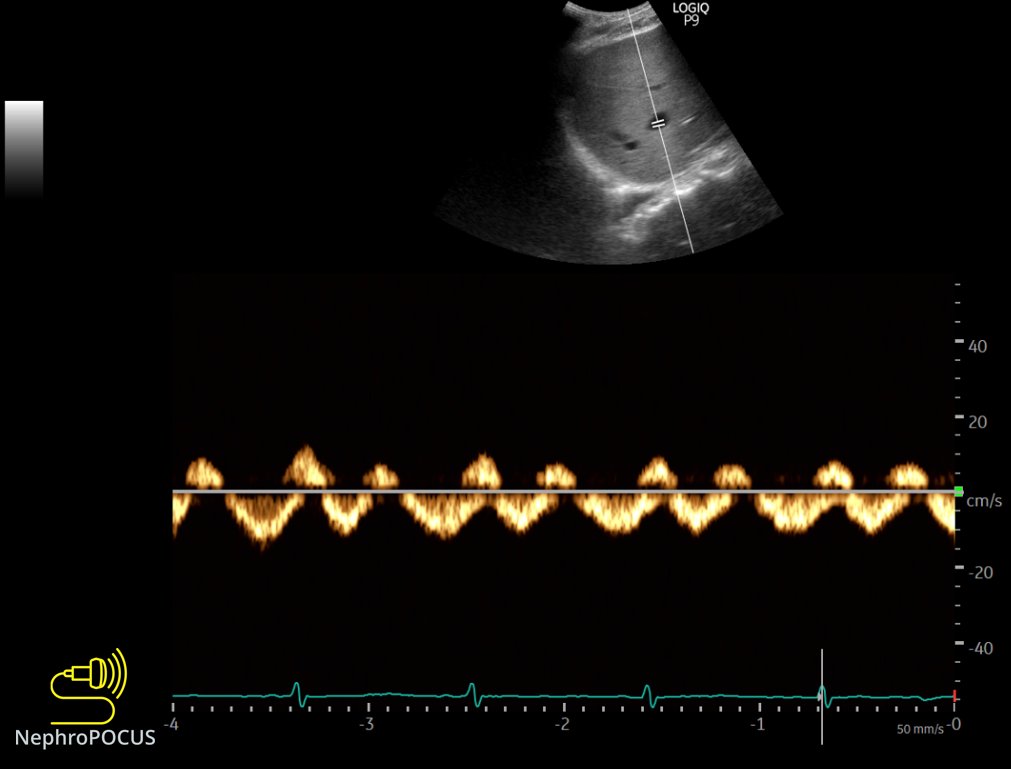
Images for #VExUS #POCUS enthusiasts
Hepatic vein https://abs.twimg.com/emoji/v2/... draggable="false" alt="👇" title="Rückhand Zeigefinger nach unten" aria-label="Emoji: Rückhand Zeigefinger nach unten">Looks almost normal
This is a kind of tracing where S and D can be confused for one another without EKG
Hepatic vein
This is a kind of tracing where S and D can be confused for one another without EKG
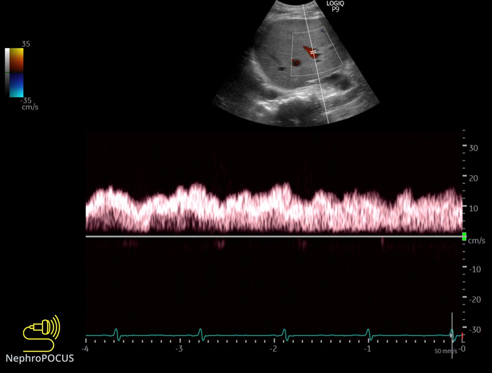
Portal #VExUS  https://abs.twimg.com/emoji/v2/... draggable="false" alt="👇" title="Rückhand Zeigefinger nach unten" aria-label="Emoji: Rückhand Zeigefinger nach unten"> - normal
Renal not checked; on dialysis for several years
Renal not checked; on dialysis for several years
PSAX with curvilinear probe. Though not of great quality, shows the circumferential nature of the effusion.
#POCUS #echofirst
#POCUS #echofirst
Apical 4 chamber #POCUS

 Read on Twitter
 Obtained during quiet breathing; no sniff" title="Mitral inflow Doppler #echofirst to assess echocardiographic pulsus paradoxus https://abs.twimg.com/emoji/v2/... draggable="false" alt="👇" title="Rückhand Zeigefinger nach unten" aria-label="Emoji: Rückhand Zeigefinger nach unten">Obtained during quiet breathing; no sniff" class="img-responsive" style="max-width:100%;"/>
 Looks almost normalThis is a kind of tracing where S and D can be confused for one another without EKG" title="Images for #VExUS #POCUS enthusiastsHepatic vein https://abs.twimg.com/emoji/v2/... draggable="false" alt="👇" title="Rückhand Zeigefinger nach unten" aria-label="Emoji: Rückhand Zeigefinger nach unten">Looks almost normalThis is a kind of tracing where S and D can be confused for one another without EKG" class="img-responsive" style="max-width:100%;"/>
 - normalRenal not checked; on dialysis for several years" title="Portal #VExUS https://abs.twimg.com/emoji/v2/... draggable="false" alt="👇" title="Rückhand Zeigefinger nach unten" aria-label="Emoji: Rückhand Zeigefinger nach unten"> - normalRenal not checked; on dialysis for several years" class="img-responsive" style="max-width:100%;"/>
