A lot of talk about #CVST Rare to have neuro in the news, so here& #39;s a quick #neurology #Tweetorial on cerebral venous sinus thrombosis.
#MedEd #MedTwitter #neurotwitter @CPSolvers @rabihmgeha @DxRxEdu #EndNeurophobia
#MedEd #MedTwitter #neurotwitter @CPSolvers @rabihmgeha @DxRxEdu #EndNeurophobia
I will focus on the condition/neurology, not the possible association w/vaccine or hematology. I leave that to experts like @shemarmoore and her excellent thread here: https://twitter.com/acweyand/status/1382156548571590657">https://twitter.com/acweyand/...
First, what are the cerebral venous sinuses?
They are the final pathway of venous drainage from the brain into the jugular veins.
They are formed from folds in the dura.
They are the final pathway of venous drainage from the brain into the jugular veins.
They are formed from folds in the dura.
The superior sagittal sinus runs in interhemispheric fissure, leads back to the transverse sinuses in tentorium, which pass by way of sigmoid sinuses into jugular veins. Straight sinus joins transverse sinuses and superior sagittal sinus at torcula (which means wine press!)
The most famous sinus is the cavernous sinus, through which CN 3, 4, 6, V1/V2, and carotid pass…I think that’s the only place in the body in which an artery passes through a vein!? right @AvrahamCooperMD ?
But cavernous sinus thrombosis is super rare https://twitter.com/AaronLBerkowitz/status/1272306877938855937">https://twitter.com/AaronLBer...
But cavernous sinus thrombosis is super rare https://twitter.com/AaronLBerkowitz/status/1272306877938855937">https://twitter.com/AaronLBer...
When I teach #neurology for the non-neurologist, I like to make medical metaphors:
Stroke = heart attack of brain
Seizure = brain arrhythmia
ICP headache worse when supine = brain orthopnea
So what& #39;s CVST? DVT of the brain!
What do you think @cpsolvers @rabihmgeha @DxRxEdu ?
Stroke = heart attack of brain
Seizure = brain arrhythmia
ICP headache worse when supine = brain orthopnea
So what& #39;s CVST? DVT of the brain!
What do you think @cpsolvers @rabihmgeha @DxRxEdu ?
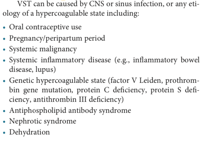
The DDX includes any cause of a hypercoaguable state (inherited, acquired, malignancy, post-partum)...
but also local processes such as head trauma, intracranial infections (meningitis, sinusitis, otitis, etc.…
see list here from https://www.amazon.com/Lange-Clinical-Neurology-Neuroanatomy-Localization-Based-ebook/dp/B01N8S6KVF">https://www.amazon.com/Lange-Cli...
but also local processes such as head trauma, intracranial infections (meningitis, sinusitis, otitis, etc.…
see list here from https://www.amazon.com/Lange-Clinical-Neurology-Neuroanatomy-Localization-Based-ebook/dp/B01N8S6KVF">https://www.amazon.com/Lange-Cli...
How does CVST present?
- Sx/signs of https://abs.twimg.com/emoji/v2/... draggable="false" alt="⬆️" title="Pfeil nach oben" aria-label="Emoji: Pfeil nach oben"> ICP: Headache, blurred vision (papilledema), double vision (from pressure on CN6), seizures; if severe
  https://abs.twimg.com/emoji/v2/... draggable="false" alt="⬇️" title="Pfeil nach unten" aria-label="Emoji: Pfeil nach unten"> level of consciousness.
- Stroke or hemorrhage with corresponding focal deficits (Cortical vein thrombosis can->convexal SAH).
- Sx/signs of
- Stroke or hemorrhage with corresponding focal deficits (Cortical vein thrombosis can->convexal SAH).
VST = a rare cause of cerebrovascular events, < 1%! One of my amazing mentors Steve Feske @harvardneuromds once said “with any stroke/hemorrhage, always ask "could this be venous?” or you’ll never remember to think of it and you’ll miss it!"
How& #39;s that for a pearl!?
How& #39;s that for a pearl!?
when to consider VST in pt w/ intracerebral hemorrhage?
A few radiology clues
- Location adjacent to a sinus
- Edema out of proportion to hemorrhage (most spontaneous ICH have relatively little edema around them)
more in GIF
other neuroradiology clues to VST @tabby_kennedy?
A few radiology clues
- Location adjacent to a sinus
- Edema out of proportion to hemorrhage (most spontaneous ICH have relatively little edema around them)
more in GIF
other neuroradiology clues to VST @tabby_kennedy?
On non con head CT look for CORD SIGN (hyperdense sinus)
If contrast given, look for empty delta sign (lack of filling at confluence of sinuses)
If contrast given, look for empty delta sign (lack of filling at confluence of sinuses)
If suspicion for CVST, get CTV or MRV and look for filling defect . If scan was already ordered without these, can look for filling defect on post-contrast sequences or clot on GRE/SWI (will be dark)
see below and https://n.neurology.org/content/82/22/e188">https://n.neurology.org/content/8...
see below and https://n.neurology.org/content/82/22/e188">https://n.neurology.org/content/8...
Treatment is LMWH or heparin acutely (even if ICH present!), dagibitran or warfarin subsequently https://jamanetwork.com/journals/jamaneurology/fullarticle/2749167
Months">https://jamanetwork.com/journals/... if provoked/temporary cause vs need for long term AC
(Note thread from @acweyand above re: complexities of mgt if concurrent coagulopathy)
Months">https://jamanetwork.com/journals/... if provoked/temporary cause vs need for long term AC
(Note thread from @acweyand above re: complexities of mgt if concurrent coagulopathy)
In severe/extreme cases, catheter based therapies may be considered. Some data reviewed in context of this clinical reasoning case from @AANMember @RoyStrowdMD section my colleagues and i wrote up when residents @harvardneuromds
https://n.neurology.org/content/neurology/82/22/e188.full.pdf">https://n.neurology.org/content/n...
(check out the images)!
https://n.neurology.org/content/neurology/82/22/e188.full.pdf">https://n.neurology.org/content/n...
(check out the images)!
Also must manage complications:
- elevated ICP
- seizures
- supportive care
@caseyalbin surely would have more to teach us about these!
- elevated ICP
- seizures
- supportive care
@caseyalbin surely would have more to teach us about these!
I hope this helps!
Please add/teach us: @caseyalbin @namorrismd @Tracey1milligan @HollyEHinson @MadSattinJ @SherryChou399 @neurocritical
Figs/text are from https://www.amazon.com/Lange-Clinical-Neurology-Neuroanatomy-Localization-Based-ebook/dp/B01N8S6KVF
https://www.amazon.com/Lange-Cli... href="https://twtext.com//hashtag/EndNeurophobia"> #EndNeurophobia https://abs.twimg.com/emoji/v2/... draggable="false" alt="🧠" title="Gehirn" aria-label="Emoji: Gehirn">
 https://abs.twimg.com/emoji/v2/... draggable="false" alt="❤️" title="Rotes Herz" aria-label="Emoji: Rotes Herz"> @MariaMjaleman @NMatch2021 @aszelikovich @gabifpucci
Please add/teach us: @caseyalbin @namorrismd @Tracey1milligan @HollyEHinson @MadSattinJ @SherryChou399 @neurocritical
Figs/text are from https://www.amazon.com/Lange-Clinical-Neurology-Neuroanatomy-Localization-Based-ebook/dp/B01N8S6KVF

 Read on Twitter


