** MONTHLY ILLUSTRATIVE CASE **
A 46-year-old male with an aortic homograft is admitted with infective endocarditis. We performed a TEE rule out complications.
#echofirst #FOAMEd #POCUS
Personal identifiers modified for confidentiality
1/
There is a large heterogeneous area of the aortic root that is suggestive of an abscess.
 https://abs.twimg.com/emoji/v2/... draggable="false" alt="😬" title="Grimasse schneidendes Gesicht" aria-label="Emoji: Grimasse schneidendes Gesicht">
2//
2//
The aortic homograft apperas well seated with only mild transvalvular regurgitation, no paravalvular regurgitation, and no aortic valve vegetation.
 https://abs.twimg.com/emoji/v2/... draggable="false" alt="😌" title="Erleichtertes Gesicht" aria-label="Emoji: Erleichtertes Gesicht">
3//
3//
But perhaps more interesting is the mitral valve.
 https://abs.twimg.com/emoji/v2/... draggable="false" alt="🧐" title="Gesicht mit Monokel" aria-label="Emoji: Gesicht mit Monokel">
4//
4//
So let& #39;s take a quick vote, how would you grade the severity of mitral regurgitation?
 https://abs.twimg.com/emoji/v2/... draggable="false" alt="🤔" title="Denkendes Gesicht" aria-label="Emoji: Denkendes Gesicht">
5//
5//
Always start a valve assessment with an 2D assessment of the structure. There is calcification of the mitral valve and subvalvular apparatus. Look closely, and a small coaptation gap is clearly visible.
6//
6//
As seen with colour Doppler, the mitral regurgitation flows through this coaptation cap into a centrally directed jet.
7//
7//
The mitral valve is well suited to 3D imaging. To orient you, we are in the LA looking down at the mitral valve, the anterior leaflet is on top and posterior leaflet below. The aortic valve sits at 12 o& #39;clock on the top face of the 3D pyramid.
8//
8//
Again applying colour Doppler, we see the mitral regurgitation originating all along the coaptation line.
9//
9//
But take a closer look at the valve mechanics. The valve appears to studder before closing. If you haven& #39;t already picked up on the issue, this is a good giveaway.
 https://abs.twimg.com/emoji/v2/... draggable="false" alt="🧐" title="Gesicht mit Monokel" aria-label="Emoji: Gesicht mit Monokel">
10//
10//
I& #39;m going to slow down a clip of 2D colour Doppler, and ask you to pay attention to the timing of the mitral regurgitation as it relates to the ECG.
11//
11//
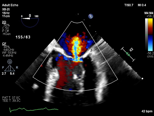
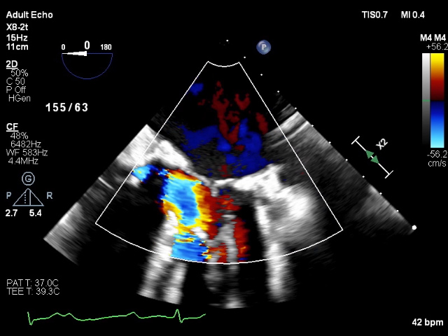
The mitral regurgitation occurs in diastole, and is absent in systole. Wot?
 https://abs.twimg.com/emoji/v2/... draggable="false" alt="🤔" title="Denkendes Gesicht" aria-label="Emoji: Denkendes Gesicht">
 https://abs.twimg.com/emoji/v2/... draggable="false" alt="🤔" title="Denkendes Gesicht" aria-label="Emoji: Denkendes Gesicht">
12//
12//
Here are some still shots to prove it!
 https://abs.twimg.com/emoji/v2/... draggable="false" alt="🤯" title="Explodierender Kopf" aria-label="Emoji: Explodierender Kopf">
13//
13//
You, my friend, have diagnosed diastolic mitral regurgitation! https://abs.twimg.com/emoji/v2/... draggable="false" alt="🎉" title="Partyknaller" aria-label="Emoji: Partyknaller">
 https://abs.twimg.com/emoji/v2/... draggable="false" alt="🎉" title="Partyknaller" aria-label="Emoji: Partyknaller">
Diastolic mitral regurgitation is rare and generally requires two criteria:
- conduction disease
- elevated LV filling pressures
14//
Diastolic mitral regurgitation is rare and generally requires two criteria:
- conduction disease
- elevated LV filling pressures
14//
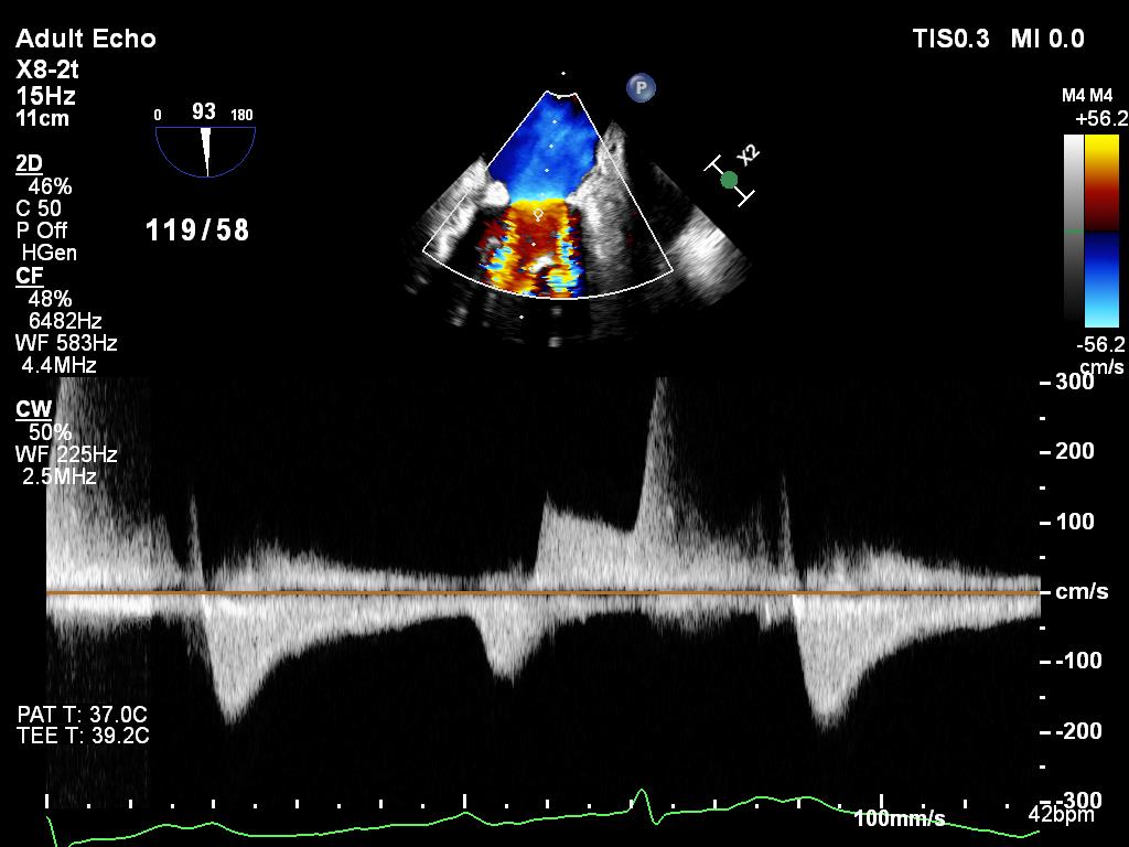
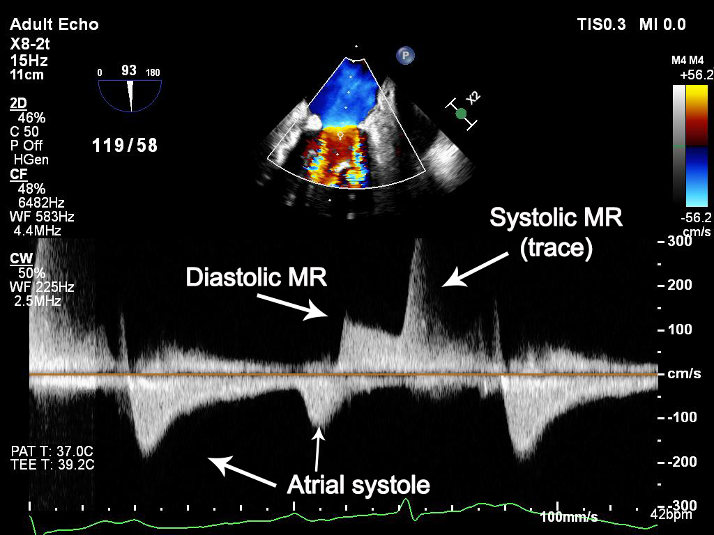
Here we see 2:1 AV conduction (a complication of the aortic root abscess). In this case, atrial systole occurs twice and there is a very long PR interval in every cardiac cycle. At a minimum, we would assume the LVEDP to approximate the LA pressure.
15//
15//
But the double-whammy is the aortic regurgitation. After atrial systole, the LVEDP continues to increase due to the aortic regurgitation. This results in the LV pressure EXCEEDING the LA pressures during diastole.
16//
16//
The mitral valve remains tethered and open in diastole. The diastolic pressure gradient drives blood from the LV to LA. Once the LV contracts and geometry changes, the MV closes!
 https://abs.twimg.com/emoji/v2/... draggable="false" alt="💡" title="Elektrische Glühbirne" aria-label="Emoji: Elektrische Glühbirne">
17//
17//
This is a rare, interesting, and dare I say super nerdy variant of function mitral regurgitation. And in this instance, is of no physiologic consequence.
18//
18//
Using this continuous wave Doppler tracing as a guide, can anyone draw what invasive hemodynamics would show?
21//
21//
As always, open to and excited for the discussion.
22//
22//
I hope you like this month& #39;s illustrative case. Follow and share if you enjoyed. More to come!
OTHER CASES:
Pericardial Waffle: https://twitter.com/OKiamanesh/status/1302045240422006785
Aortic">https://twitter.com/OKiamanes... Thrombus: https://twitter.com/OKiamanesh/status/1287536558632046592
Constriction:">https://twitter.com/OKiamanes... https://twitter.com/OKiamanesh/status/1267632487406219265
23//">https://twitter.com/OKiamanes...

 Read on Twitter
 13//" title="Here are some still shots to prove it!https://abs.twimg.com/emoji/v2/... draggable="false" alt="🤯" title="Explodierender Kopf" aria-label="Emoji: Explodierender Kopf">13//">
 13//" title="Here are some still shots to prove it!https://abs.twimg.com/emoji/v2/... draggable="false" alt="🤯" title="Explodierender Kopf" aria-label="Emoji: Explodierender Kopf">13//">


