Holiday snaps No.4
Arm
@ZHOUSEH @MattieFarzin @rennmont @DrJPLow @RunjanChetty @ariella8 @JMGardnerMD @01sth02 @TheKarenPinto @MAHoureih @DrBMcGinn @s_prendeville @DrGeeONE @Divya_Madhala @pembeoltulu @Meghna0630 @Mattzaborowski1
#BSTpath #PathTwitter #dermpath #SurgPath
Arm
@ZHOUSEH @MattieFarzin @rennmont @DrJPLow @RunjanChetty @ariella8 @JMGardnerMD @01sth02 @TheKarenPinto @MAHoureih @DrBMcGinn @s_prendeville @DrGeeONE @Divya_Madhala @pembeoltulu @Meghna0630 @Mattzaborowski1
#BSTpath #PathTwitter #dermpath #SurgPath
Diagnosis:
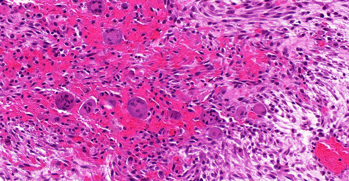
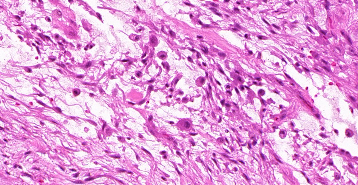
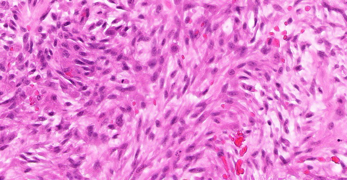
Nodular fasciitis
Nodular fasciitis
1/ Once regarded as a reactive lesion, this pseudosarcomatous is a benign neoplasm that are self-limiting.
2/ They usually present as a rapid growing lesion and only ~50% of patients report a previous associated trauma. https://pubmed.ncbi.nlm.nih.gov/6462780/ ">https://pubmed.ncbi.nlm.nih.gov/6462780/&...
2/ They usually present as a rapid growing lesion and only ~50% of patients report a previous associated trauma. https://pubmed.ncbi.nlm.nih.gov/6462780/ ">https://pubmed.ncbi.nlm.nih.gov/6462780/&...
3/These lesions are circumscribed & usually less than 3cm.
The rule of thumb https://abs.twimg.com/emoji/v2/... draggable="false" alt="➡️" title="Pfeil nach rechts" aria-label="Emoji: Pfeil nach rechts"> Whole lesion should fit on one slide!
4/More common in upper extremities, but it can occur at almost any anatomical site, including intravascular forms and in any age. https://pubmed.ncbi.nlm.nih.gov/7246849/ ">https://pubmed.ncbi.nlm.nih.gov/7246849/&...
The rule of thumb
4/More common in upper extremities, but it can occur at almost any anatomical site, including intravascular forms and in any age. https://pubmed.ncbi.nlm.nih.gov/7246849/ ">https://pubmed.ncbi.nlm.nih.gov/7246849/&...
5/ They have a characteristic gene fusion MYH9-USP. This can be useful in unusual cases (eg large lesions).
6/ USP6 fusions can also be found in aneurysmal bone cysts and myositis ossificans (MO). https://pubmed.ncbi.nlm.nih.gov/28752842/ ">https://pubmed.ncbi.nlm.nih.gov/28752842/...
6/ USP6 fusions can also be found in aneurysmal bone cysts and myositis ossificans (MO). https://pubmed.ncbi.nlm.nih.gov/28752842/ ">https://pubmed.ncbi.nlm.nih.gov/28752842/...
7/Classic features are:
 https://abs.twimg.com/emoji/v2/... draggable="false" alt="☑️" title="Kästchen mit Häkchen" aria-label="Emoji: Kästchen mit Häkchen">Myxoid or collagenous matrix
 https://abs.twimg.com/emoji/v2/... draggable="false" alt="☑️" title="Kästchen mit Häkchen" aria-label="Emoji: Kästchen mit Häkchen">Cellular myo/fibroblastic cells with plump nuclei & occasional nucleoli
 https://abs.twimg.com/emoji/v2/... draggable="false" alt="☑️" title="Kästchen mit Häkchen" aria-label="Emoji: Kästchen mit Häkchen">Tissue culture-like cells
 https://abs.twimg.com/emoji/v2/... draggable="false" alt="☑️" title="Kästchen mit Häkchen" aria-label="Emoji: Kästchen mit Häkchen">S/C-shaped fascicles, sometimes storiform
 https://abs.twimg.com/emoji/v2/... draggable="false" alt="☑️" title="Kästchen mit Häkchen" aria-label="Emoji: Kästchen mit Häkchen">RBC extravasation
 https://abs.twimg.com/emoji/v2/... draggable="false" alt="☑️" title="Kästchen mit Häkchen" aria-label="Emoji: Kästchen mit Häkchen">Lymphocytes
 https://abs.twimg.com/emoji/v2/... draggable="false" alt="❌" title="Kreuzzeichen" aria-label="Emoji: Kreuzzeichen">Hyperchromatic/pleomorphic cells
8/ But also can have:
 https://abs.twimg.com/emoji/v2/... draggable="false" alt="☑️" title="Kästchen mit Häkchen" aria-label="Emoji: Kästchen mit Häkchen"> Numerous mitoses
 https://abs.twimg.com/emoji/v2/... draggable="false" alt="☑️" title="Kästchen mit Häkchen" aria-label="Emoji: Kästchen mit Häkchen"> Scattered giant cells
 https://abs.twimg.com/emoji/v2/... draggable="false" alt="☑️" title="Kästchen mit Häkchen" aria-label="Emoji: Kästchen mit Häkchen"> Focal metaplastic bone formation (relationship to MO)
 https://abs.twimg.com/emoji/v2/... draggable="false" alt="☑️" title="Kästchen mit Häkchen" aria-label="Emoji: Kästchen mit Häkchen"> Centripetally oriented capillaries
 https://abs.twimg.com/emoji/v2/... draggable="false" alt="☑️" title="Kästchen mit Häkchen" aria-label="Emoji: Kästchen mit Häkchen"> Cystic change

 Read on Twitter
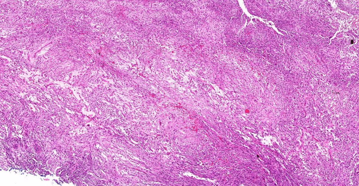
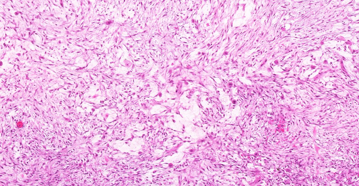
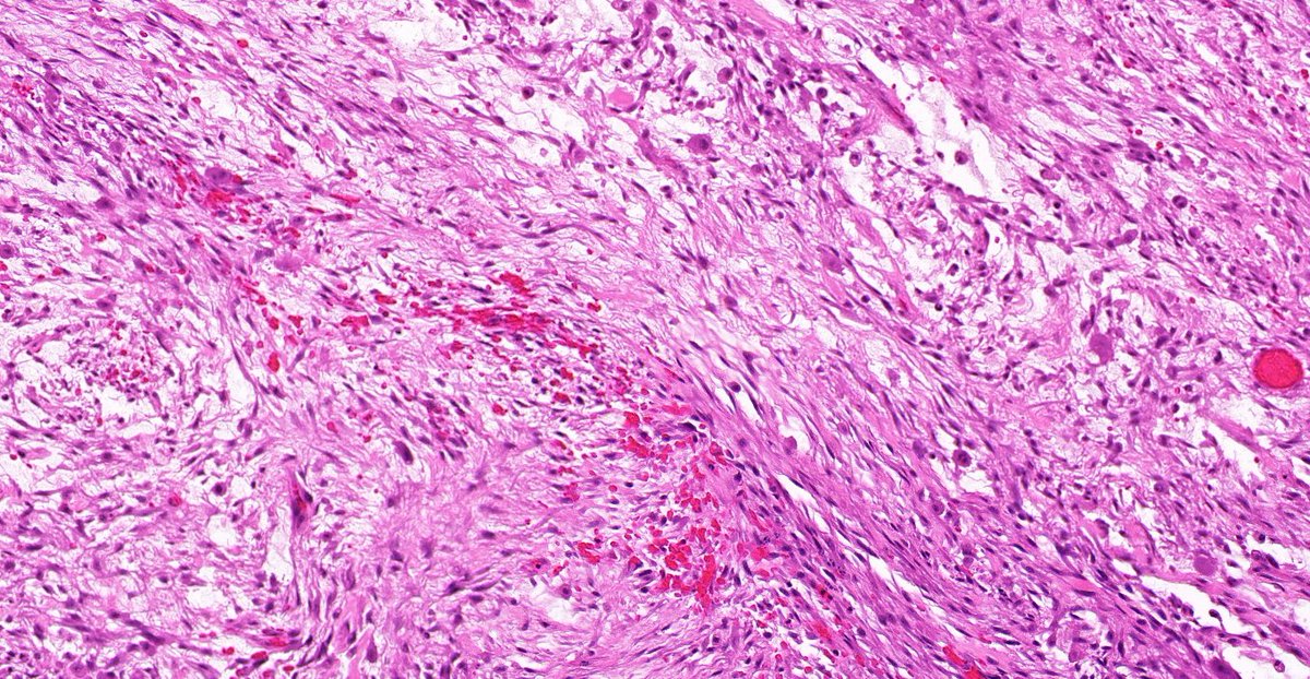
 Myxoid or collagenous matrixhttps://abs.twimg.com/emoji/v2/... draggable="false" alt="☑️" title="Kästchen mit Häkchen" aria-label="Emoji: Kästchen mit Häkchen">Cellular myo/fibroblastic cells with plump nuclei & occasional nucleolihttps://abs.twimg.com/emoji/v2/... draggable="false" alt="☑️" title="Kästchen mit Häkchen" aria-label="Emoji: Kästchen mit Häkchen">Tissue culture-like cellshttps://abs.twimg.com/emoji/v2/... draggable="false" alt="☑️" title="Kästchen mit Häkchen" aria-label="Emoji: Kästchen mit Häkchen">S/C-shaped fascicles, sometimes storiformhttps://abs.twimg.com/emoji/v2/... draggable="false" alt="☑️" title="Kästchen mit Häkchen" aria-label="Emoji: Kästchen mit Häkchen">RBC extravasationhttps://abs.twimg.com/emoji/v2/... draggable="false" alt="☑️" title="Kästchen mit Häkchen" aria-label="Emoji: Kästchen mit Häkchen">Lymphocyteshttps://abs.twimg.com/emoji/v2/... draggable="false" alt="❌" title="Kreuzzeichen" aria-label="Emoji: Kreuzzeichen">Hyperchromatic/pleomorphic cells" title="7/Classic features are:https://abs.twimg.com/emoji/v2/... draggable="false" alt="☑️" title="Kästchen mit Häkchen" aria-label="Emoji: Kästchen mit Häkchen">Myxoid or collagenous matrixhttps://abs.twimg.com/emoji/v2/... draggable="false" alt="☑️" title="Kästchen mit Häkchen" aria-label="Emoji: Kästchen mit Häkchen">Cellular myo/fibroblastic cells with plump nuclei & occasional nucleolihttps://abs.twimg.com/emoji/v2/... draggable="false" alt="☑️" title="Kästchen mit Häkchen" aria-label="Emoji: Kästchen mit Häkchen">Tissue culture-like cellshttps://abs.twimg.com/emoji/v2/... draggable="false" alt="☑️" title="Kästchen mit Häkchen" aria-label="Emoji: Kästchen mit Häkchen">S/C-shaped fascicles, sometimes storiformhttps://abs.twimg.com/emoji/v2/... draggable="false" alt="☑️" title="Kästchen mit Häkchen" aria-label="Emoji: Kästchen mit Häkchen">RBC extravasationhttps://abs.twimg.com/emoji/v2/... draggable="false" alt="☑️" title="Kästchen mit Häkchen" aria-label="Emoji: Kästchen mit Häkchen">Lymphocyteshttps://abs.twimg.com/emoji/v2/... draggable="false" alt="❌" title="Kreuzzeichen" aria-label="Emoji: Kreuzzeichen">Hyperchromatic/pleomorphic cells" class="img-responsive" style="max-width:100%;"/>
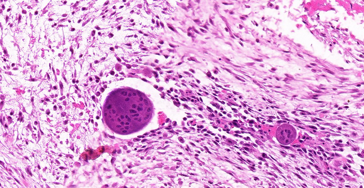 Numerous mitoseshttps://abs.twimg.com/emoji/v2/... draggable="false" alt="☑️" title="Kästchen mit Häkchen" aria-label="Emoji: Kästchen mit Häkchen"> Scattered giant cellshttps://abs.twimg.com/emoji/v2/... draggable="false" alt="☑️" title="Kästchen mit Häkchen" aria-label="Emoji: Kästchen mit Häkchen"> Focal metaplastic bone formation (relationship to MO)https://abs.twimg.com/emoji/v2/... draggable="false" alt="☑️" title="Kästchen mit Häkchen" aria-label="Emoji: Kästchen mit Häkchen"> Centripetally oriented capillarieshttps://abs.twimg.com/emoji/v2/... draggable="false" alt="☑️" title="Kästchen mit Häkchen" aria-label="Emoji: Kästchen mit Häkchen"> Cystic change" title="8/ But also can have:https://abs.twimg.com/emoji/v2/... draggable="false" alt="☑️" title="Kästchen mit Häkchen" aria-label="Emoji: Kästchen mit Häkchen"> Numerous mitoseshttps://abs.twimg.com/emoji/v2/... draggable="false" alt="☑️" title="Kästchen mit Häkchen" aria-label="Emoji: Kästchen mit Häkchen"> Scattered giant cellshttps://abs.twimg.com/emoji/v2/... draggable="false" alt="☑️" title="Kästchen mit Häkchen" aria-label="Emoji: Kästchen mit Häkchen"> Focal metaplastic bone formation (relationship to MO)https://abs.twimg.com/emoji/v2/... draggable="false" alt="☑️" title="Kästchen mit Häkchen" aria-label="Emoji: Kästchen mit Häkchen"> Centripetally oriented capillarieshttps://abs.twimg.com/emoji/v2/... draggable="false" alt="☑️" title="Kästchen mit Häkchen" aria-label="Emoji: Kästchen mit Häkchen"> Cystic change">
 Numerous mitoseshttps://abs.twimg.com/emoji/v2/... draggable="false" alt="☑️" title="Kästchen mit Häkchen" aria-label="Emoji: Kästchen mit Häkchen"> Scattered giant cellshttps://abs.twimg.com/emoji/v2/... draggable="false" alt="☑️" title="Kästchen mit Häkchen" aria-label="Emoji: Kästchen mit Häkchen"> Focal metaplastic bone formation (relationship to MO)https://abs.twimg.com/emoji/v2/... draggable="false" alt="☑️" title="Kästchen mit Häkchen" aria-label="Emoji: Kästchen mit Häkchen"> Centripetally oriented capillarieshttps://abs.twimg.com/emoji/v2/... draggable="false" alt="☑️" title="Kästchen mit Häkchen" aria-label="Emoji: Kästchen mit Häkchen"> Cystic change" title="8/ But also can have:https://abs.twimg.com/emoji/v2/... draggable="false" alt="☑️" title="Kästchen mit Häkchen" aria-label="Emoji: Kästchen mit Häkchen"> Numerous mitoseshttps://abs.twimg.com/emoji/v2/... draggable="false" alt="☑️" title="Kästchen mit Häkchen" aria-label="Emoji: Kästchen mit Häkchen"> Scattered giant cellshttps://abs.twimg.com/emoji/v2/... draggable="false" alt="☑️" title="Kästchen mit Häkchen" aria-label="Emoji: Kästchen mit Häkchen"> Focal metaplastic bone formation (relationship to MO)https://abs.twimg.com/emoji/v2/... draggable="false" alt="☑️" title="Kästchen mit Häkchen" aria-label="Emoji: Kästchen mit Häkchen"> Centripetally oriented capillarieshttps://abs.twimg.com/emoji/v2/... draggable="false" alt="☑️" title="Kästchen mit Häkchen" aria-label="Emoji: Kästchen mit Häkchen"> Cystic change">
 Numerous mitoseshttps://abs.twimg.com/emoji/v2/... draggable="false" alt="☑️" title="Kästchen mit Häkchen" aria-label="Emoji: Kästchen mit Häkchen"> Scattered giant cellshttps://abs.twimg.com/emoji/v2/... draggable="false" alt="☑️" title="Kästchen mit Häkchen" aria-label="Emoji: Kästchen mit Häkchen"> Focal metaplastic bone formation (relationship to MO)https://abs.twimg.com/emoji/v2/... draggable="false" alt="☑️" title="Kästchen mit Häkchen" aria-label="Emoji: Kästchen mit Häkchen"> Centripetally oriented capillarieshttps://abs.twimg.com/emoji/v2/... draggable="false" alt="☑️" title="Kästchen mit Häkchen" aria-label="Emoji: Kästchen mit Häkchen"> Cystic change" title="8/ But also can have:https://abs.twimg.com/emoji/v2/... draggable="false" alt="☑️" title="Kästchen mit Häkchen" aria-label="Emoji: Kästchen mit Häkchen"> Numerous mitoseshttps://abs.twimg.com/emoji/v2/... draggable="false" alt="☑️" title="Kästchen mit Häkchen" aria-label="Emoji: Kästchen mit Häkchen"> Scattered giant cellshttps://abs.twimg.com/emoji/v2/... draggable="false" alt="☑️" title="Kästchen mit Häkchen" aria-label="Emoji: Kästchen mit Häkchen"> Focal metaplastic bone formation (relationship to MO)https://abs.twimg.com/emoji/v2/... draggable="false" alt="☑️" title="Kästchen mit Häkchen" aria-label="Emoji: Kästchen mit Häkchen"> Centripetally oriented capillarieshttps://abs.twimg.com/emoji/v2/... draggable="false" alt="☑️" title="Kästchen mit Häkchen" aria-label="Emoji: Kästchen mit Häkchen"> Cystic change">
 Numerous mitoseshttps://abs.twimg.com/emoji/v2/... draggable="false" alt="☑️" title="Kästchen mit Häkchen" aria-label="Emoji: Kästchen mit Häkchen"> Scattered giant cellshttps://abs.twimg.com/emoji/v2/... draggable="false" alt="☑️" title="Kästchen mit Häkchen" aria-label="Emoji: Kästchen mit Häkchen"> Focal metaplastic bone formation (relationship to MO)https://abs.twimg.com/emoji/v2/... draggable="false" alt="☑️" title="Kästchen mit Häkchen" aria-label="Emoji: Kästchen mit Häkchen"> Centripetally oriented capillarieshttps://abs.twimg.com/emoji/v2/... draggable="false" alt="☑️" title="Kästchen mit Häkchen" aria-label="Emoji: Kästchen mit Häkchen"> Cystic change" title="8/ But also can have:https://abs.twimg.com/emoji/v2/... draggable="false" alt="☑️" title="Kästchen mit Häkchen" aria-label="Emoji: Kästchen mit Häkchen"> Numerous mitoseshttps://abs.twimg.com/emoji/v2/... draggable="false" alt="☑️" title="Kästchen mit Häkchen" aria-label="Emoji: Kästchen mit Häkchen"> Scattered giant cellshttps://abs.twimg.com/emoji/v2/... draggable="false" alt="☑️" title="Kästchen mit Häkchen" aria-label="Emoji: Kästchen mit Häkchen"> Focal metaplastic bone formation (relationship to MO)https://abs.twimg.com/emoji/v2/... draggable="false" alt="☑️" title="Kästchen mit Häkchen" aria-label="Emoji: Kästchen mit Häkchen"> Centripetally oriented capillarieshttps://abs.twimg.com/emoji/v2/... draggable="false" alt="☑️" title="Kästchen mit Häkchen" aria-label="Emoji: Kästchen mit Häkchen"> Cystic change">


