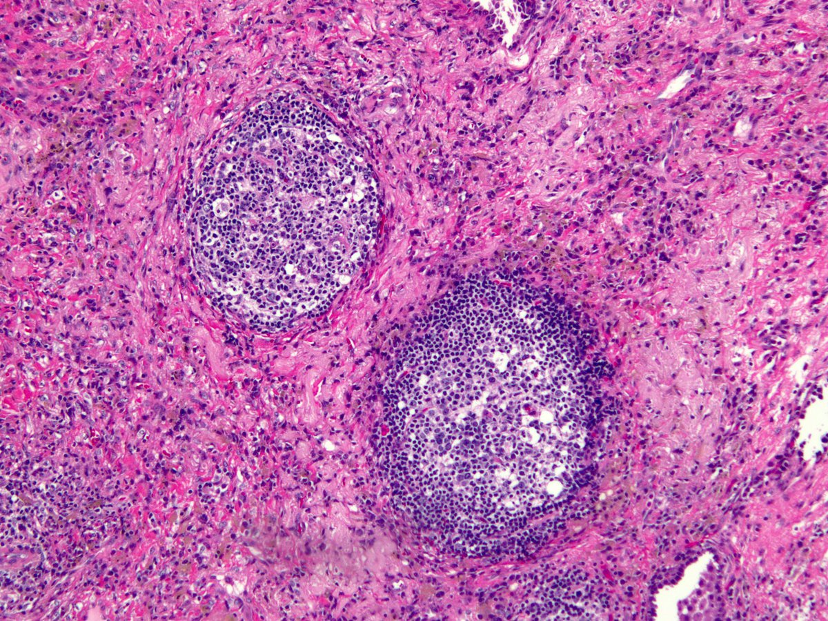
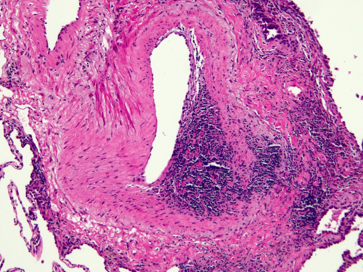
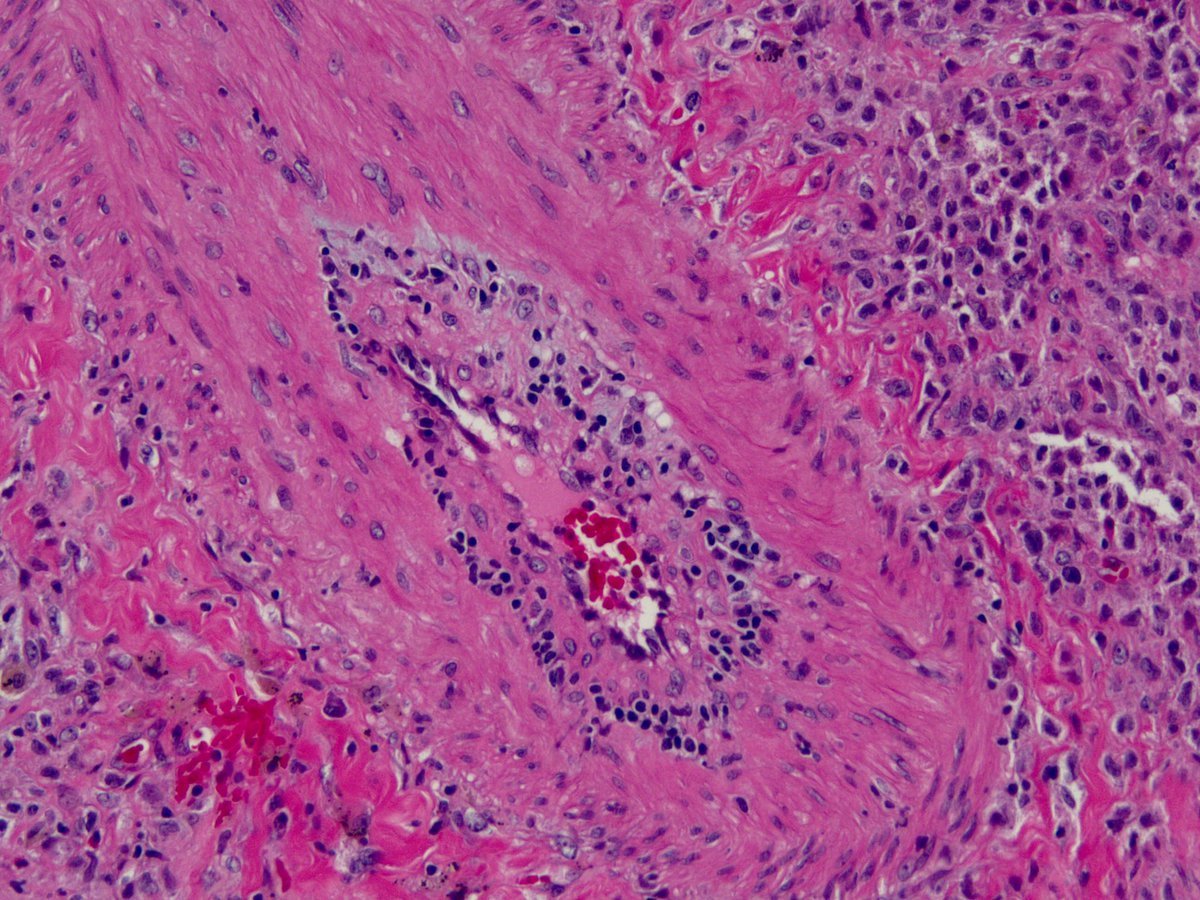
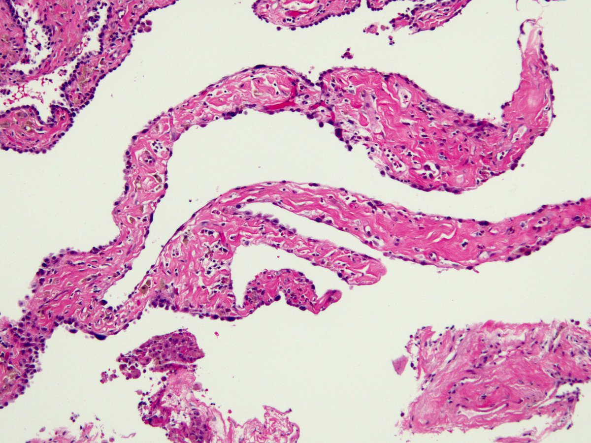
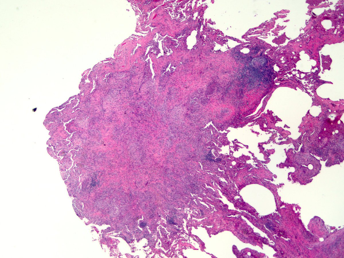
O Brave and Wise, teach me how to correlate these lung biopsy findings with clinical findings. Germinal center (pattern?) + vasculitis (uh...pattern?) + NSIP (pattern!) = ?
Clinical correlation recommended?
Some say say germinal centers are diagnostic of CTD. What say you?
Clinical correlation recommended?
Some say say germinal centers are diagnostic of CTD. What say you?
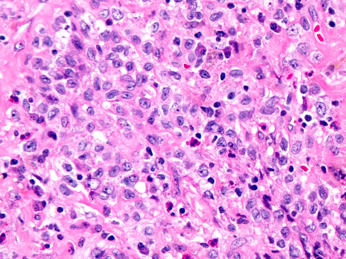
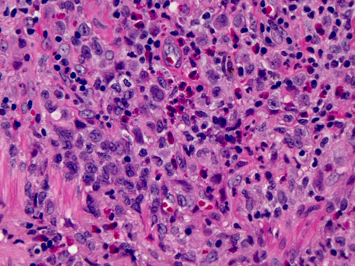
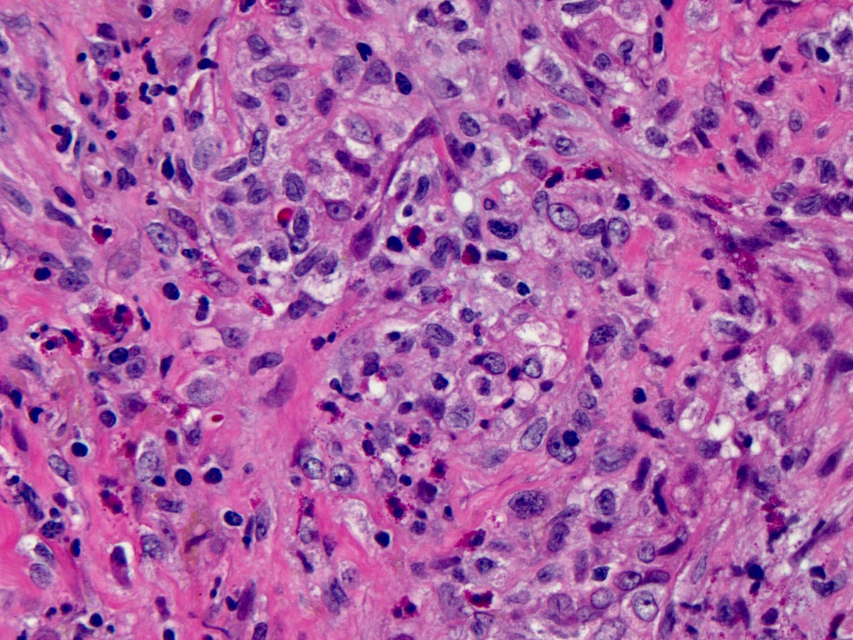
This is classic pulmonary Langerhans cell histiocytosis:
1. mm-size stellate nodules, sheets of Langerhans cells, eos
2. All the findings I posted before are distractors.
3. “Vasculitis” is common in PLCH, can look much worse than this
4. The “NSIP” pic is SRIF (needs skill)
1. mm-size stellate nodules, sheets of Langerhans cells, eos
2. All the findings I posted before are distractors.
3. “Vasculitis” is common in PLCH, can look much worse than this
4. The “NSIP” pic is SRIF (needs skill)
5. Germinal centers are entirely nonspecific. You diagnose CTD on that basis at your peril.
6. Clinicians are taught that PLCH = cystic lung dz + nodules but many patients have nodules ONLY (need pathology to diagnose these)
7. This patient needed a biopsy, is doing great
6. Clinicians are taught that PLCH = cystic lung dz + nodules but many patients have nodules ONLY (need pathology to diagnose these)
7. This patient needed a biopsy, is doing great
8. The imaging differential in these cases is metastatic cancer (h/o lung cancer in this case) and miliary infection
9. Sometimes pathology is pathognomonic but only if you know what to look for and what to ignore @IPFdoc @BDSouthern @leticiakawano https://abs.twimg.com/emoji/v2/... draggable="false" alt="😊" title="Lächelndes Gesicht mit lächelnden Augen" aria-label="Emoji: Lächelndes Gesicht mit lächelnden Augen">
 https://abs.twimg.com/emoji/v2/... draggable="false" alt="🙏🏾" title="Folded hands (durchschnittlich dunkler Hautton)" aria-label="Emoji: Folded hands (durchschnittlich dunkler Hautton)">
—————-end thread——————
9. Sometimes pathology is pathognomonic but only if you know what to look for and what to ignore @IPFdoc @BDSouthern @leticiakawano
—————-end thread——————

 Read on Twitter