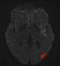
A #stroke #tweetorial. Inferior division MCA infarct often gets mistaken for PCA territory. Sometimes it can be quite difficult to distinguish MCA vs PCA territory infarcts (especially near the borderzone). #neurotwitter #medtwitter #medstudenttwitter
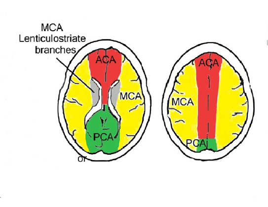
1/ Reminder of topography: MCA (yellow) and PCA (green) territory. Inferior division MCA (near the borderzone of PCA) involves the occipital lobe
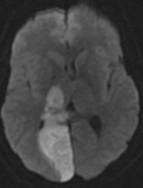
2/ This is an example of a full PCA territory infarct on MRI DWI in a patient with a PCA occlusion. PCA infarct affects the very medial occipital lobe. In a very proximal PCA occlusion, thalamus/midbrain can also be affected.
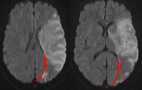
3/ This is an example of a patchy MCA territory infarct. Note that the inferior division MCA affects the partieto-occipital lobe (except for the very medial portion of the occipital lobe, which we already stated is PCA). Red line indicates the borderzone between MCA/PCA
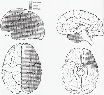
6/ Also keep in mind, the territory maps show you averages, and there is variation in individuals on the territory distributions. The image below denotes maximum, usual, and minimum territory for MCA (and there can be quite a bit of variability in the spread)
4/ Why does it matter? Confusing the territory of the infarct can mislead the clinician to the wrong mechanism/subtype. Also, infarct territory is relevant for purposes of designating a carotid stenosis symptomatic (which requires urgent intervention) vs asymptomatic stenosis.

 Read on Twitter