We know that loss of cerebral blood flow leads to changes in cellular physiology, which results in changes to the EEG brainwaves. But have you ever seen these changes occur live on your screen? A #twEEGtorial on #EEG and #ischemic injury
1/
1/
This 80yo man with AFib had been off anticoagulation for an upcoming surgery and was admitted with LKN > 24h ago with this CT head:
2/
2/
Due to encephalopathy, he was placed on cEEG. This was his baseline recording [LFF 1 Hz, HFF 70 Hz, notch 60 Hz on, S = 7 uV/mm, t = 30mm/sec]. Appreciate the left hemispheric slowing (red arrow pointing to a more obvious portion) and relative attenuation (blue arrow)
3/
3/
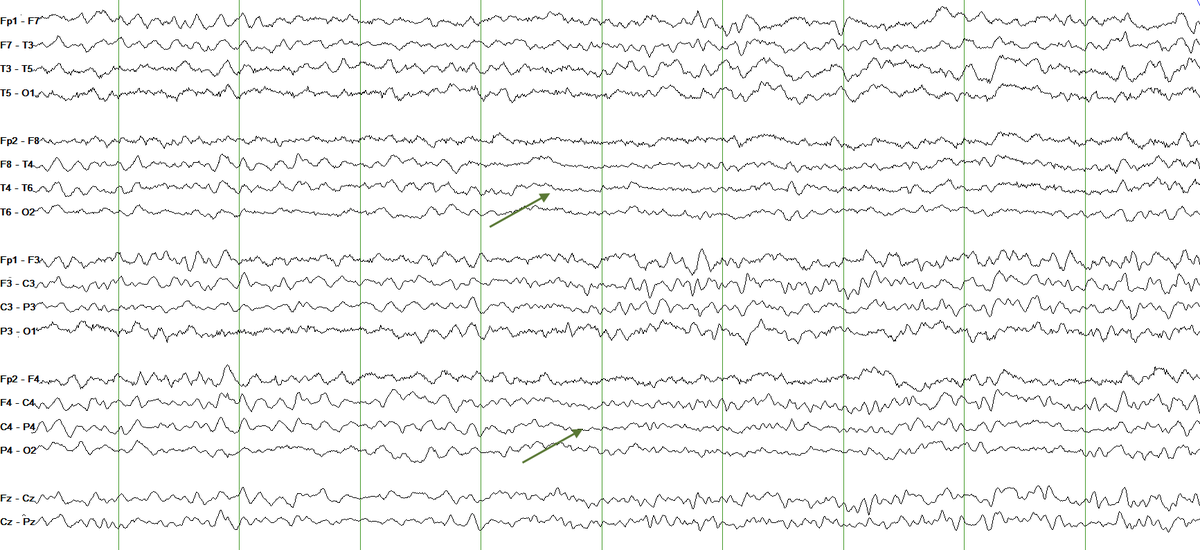
About an hour into the study, you start seeing some changes on the right side. You can appreciate more prominent theta slowing, a loss of faster frequencies and even some attenuation (green arrow pointing to a more obvious portion).
4/
4/
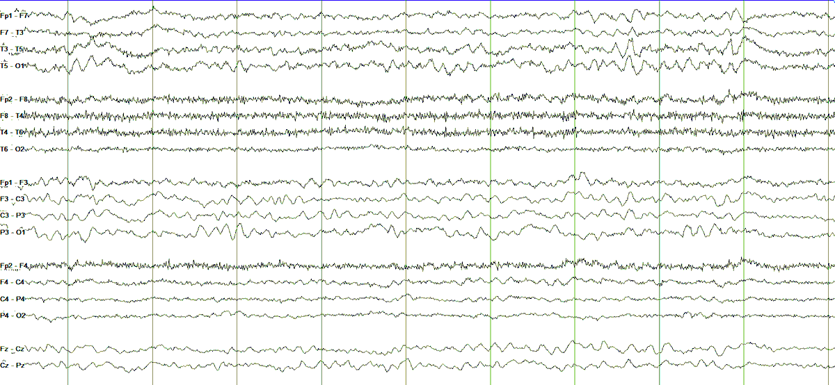
Two more hours have now elapsed. Look at that right hemisphere: very clearly attenuated. The qEEG dramatically shows a loss of power across all frequencies (pink rectangle) and the asymmetry spectrogram favors left side (blue color dominates throughout)
5/
5/
These two snapshots show the jump from a presumed reversible state (ischemia) with more prominent slowing and loss of faster frequencies to irreversible damage (infarction) with frank suppression of activity. By now, the Na/K pump has failed and there is cell death.
6/
6/
Let& #39;s confirm on imaging with a repeat CT head. Pay attention to the right hemisphere (left side of the image) and compare it with his baseline scan.
7/
7/
Here& #39;s an MRI diffusion sequence. The pump has failed and water has accumulated intracellularly causing that very bright signal (diffusion restriction), pointing to an acute ischemic stroke.
8/
8/
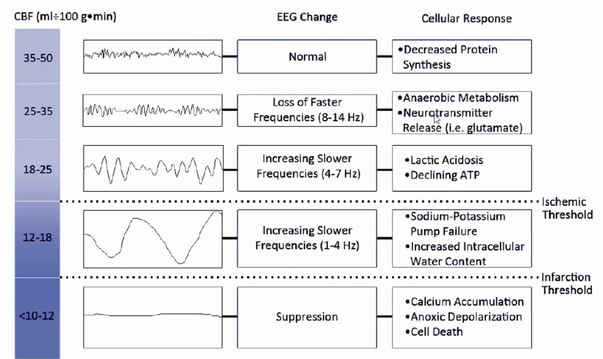
I like this table from Foreman & Claassen in Critical Care 2012 which summarizes the cellular and EEG changes that occur with progressive loss of cerebral blood flow.
9/
9/
Definitely one of those things you never want to witness, but an important reminder that EEG reflects cellular activity. Here& #39;s the link to another thread where we discussed the EEG correlates of underlying cellular (epileptogenic) activity: https://twitter.com/e_gleich/status/1295834391957114880?s=20
10/10">https://twitter.com/e_gleich/...
10/10">https://twitter.com/e_gleich/...

 Read on Twitter
![Due to encephalopathy, he was placed on cEEG. This was his baseline recording [LFF 1 Hz, HFF 70 Hz, notch 60 Hz on, S = 7 uV/mm, t = 30mm/sec]. Appreciate the left hemispheric slowing (red arrow pointing to a more obvious portion) and relative attenuation (blue arrow)3/ Due to encephalopathy, he was placed on cEEG. This was his baseline recording [LFF 1 Hz, HFF 70 Hz, notch 60 Hz on, S = 7 uV/mm, t = 30mm/sec]. Appreciate the left hemispheric slowing (red arrow pointing to a more obvious portion) and relative attenuation (blue arrow)3/](https://pbs.twimg.com/media/Egw0-rfWkAA4wgc.png)


