Catch those DVTs Early... BEFORE they get to the  https://abs.twimg.com/emoji/v2/... draggable="false" alt="💔" title="Gebrochenes Herz" aria-label="Emoji: Gebrochenes Herz"> +
  https://abs.twimg.com/emoji/v2/... draggable="false" alt="🫁" title="Lungs" aria-label="Emoji: Lungs">! #POCUS
 https://abs.twimg.com/emoji/v2/... draggable="false" alt="1⃣" title="Tastenkappe Ziffer 1" aria-label="Emoji: Tastenkappe Ziffer 1">Learn how to Easily do DVT Ultrasound
 https://abs.twimg.com/emoji/v2/... draggable="false" alt="2⃣" title="Tastenkappe Ziffer 2" aria-label="Emoji: Tastenkappe Ziffer 2">Learn how to use Compression Properly
 https://abs.twimg.com/emoji/v2/... draggable="false" alt="3⃣" title="Tastenkappe Ziffer 3" aria-label="Emoji: Tastenkappe Ziffer 3">Recognize False Positives for DVT
 https://abs.twimg.com/emoji/v2/... draggable="false" alt="✅" title="Fettes weißes Häkchen" aria-label="Emoji: Fettes weißes Häkchen">New Blog Post
  https://abs.twimg.com/emoji/v2/... draggable="false" alt="👉" title="Rückhand Zeigefinger nach rechts" aria-label="Emoji: Rückhand Zeigefinger nach rechts">
 https://abs.twimg.com/emoji/v2/... draggable="false" alt="🔗" title="Link Symbol" aria-label="Emoji: Link Symbol"> https://pocus101.com/dvt
https://pocus101.com/dvt"... href="https://twtext.com//hashtag/medtweetorial"> #medtweetorial https://abs.twimg.com/emoji/v2/... draggable="false" alt="👇" title="Rückhand Zeigefinger nach unten" aria-label="Emoji: Rückhand Zeigefinger nach unten">(1/20)
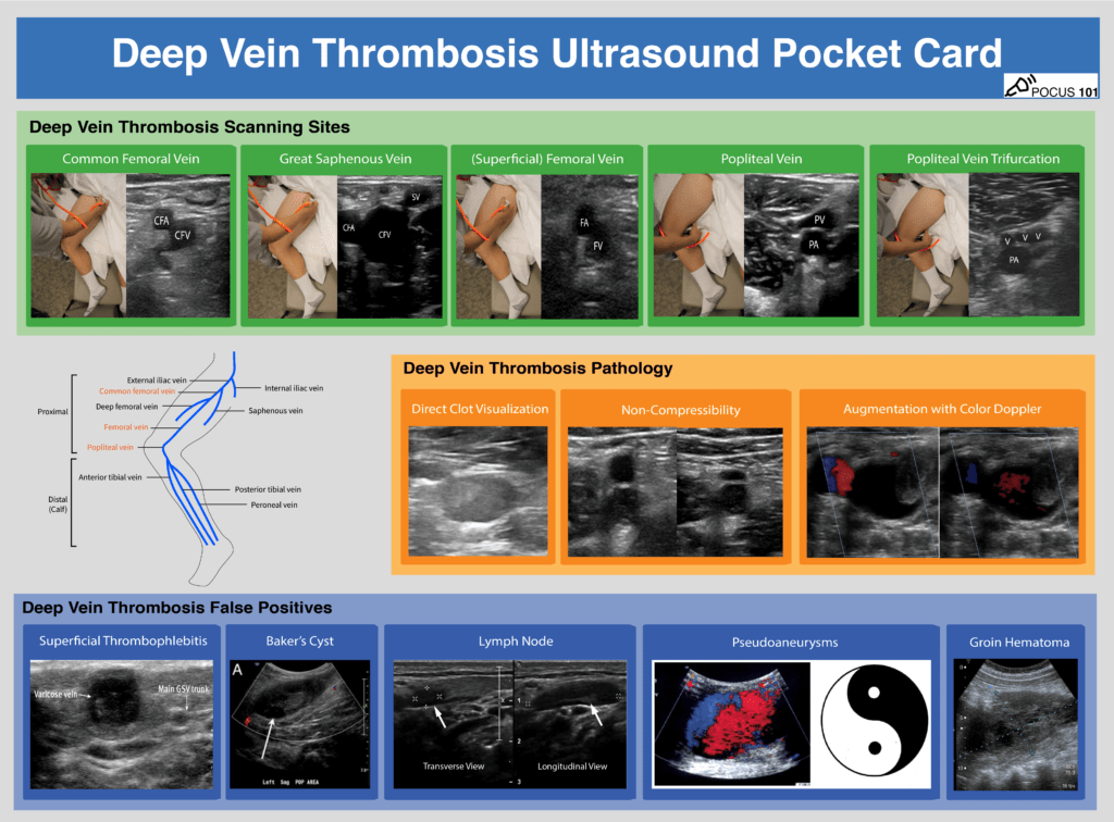
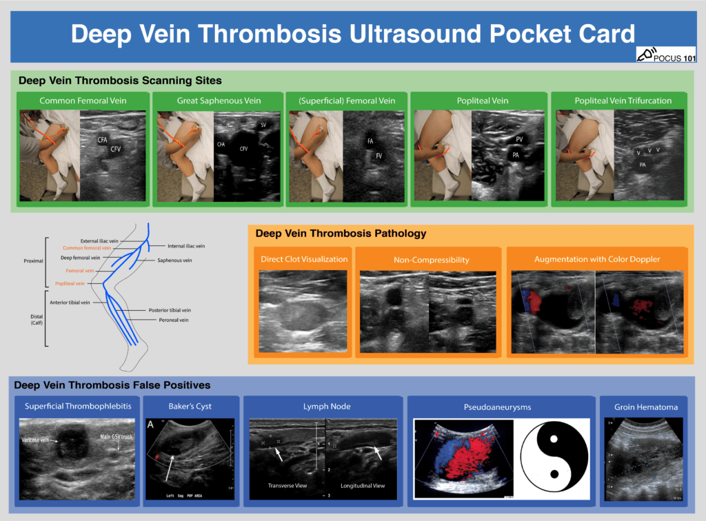
2 Download the FREE DVT Ultrasound PDF Pocket Card!
 https://abs.twimg.com/emoji/v2/... draggable="false" alt="👉" title="Rückhand Zeigefinger nach rechts" aria-label="Emoji: Rückhand Zeigefinger nach rechts">
 https://abs.twimg.com/emoji/v2/... draggable="false" alt="🔗" title="Link Symbol" aria-label="Emoji: Link Symbol"> https://pocus101.com/dvt ">https://pocus101.com/dvt"...
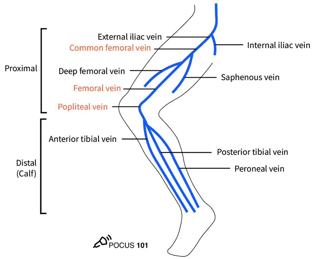
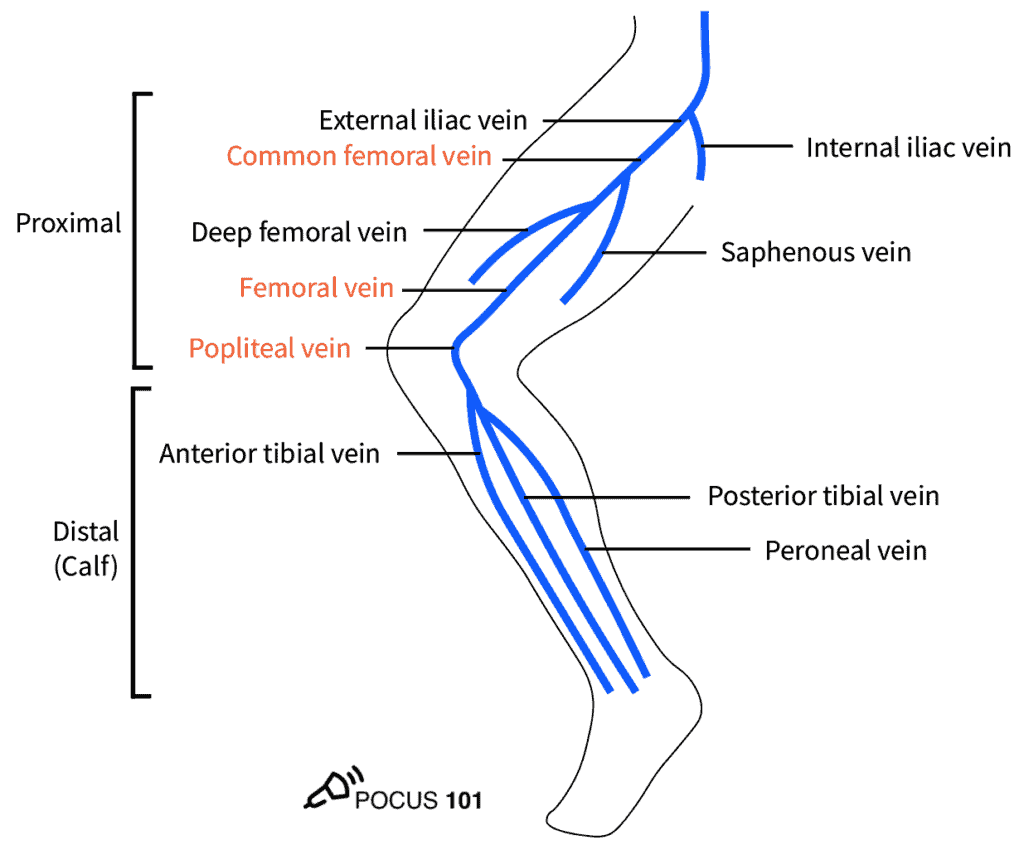
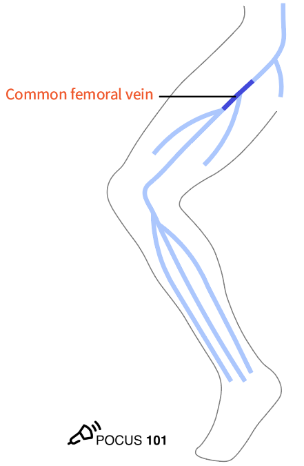
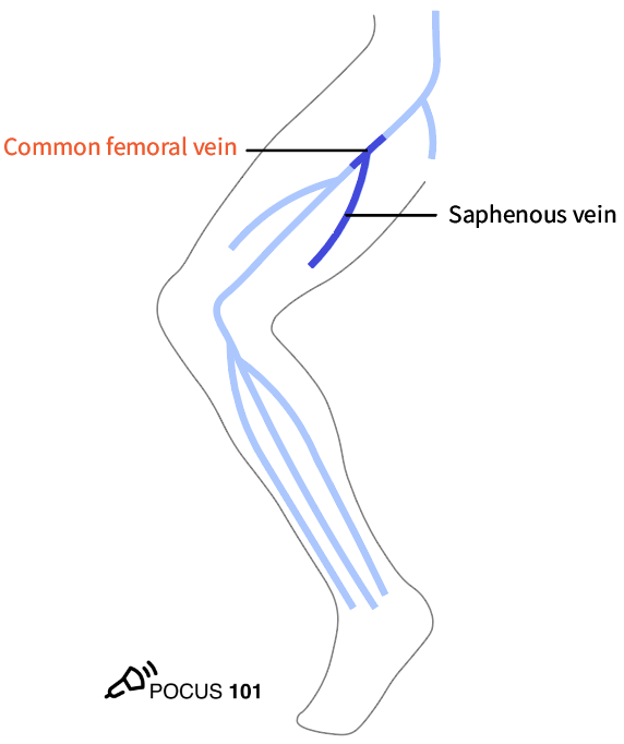
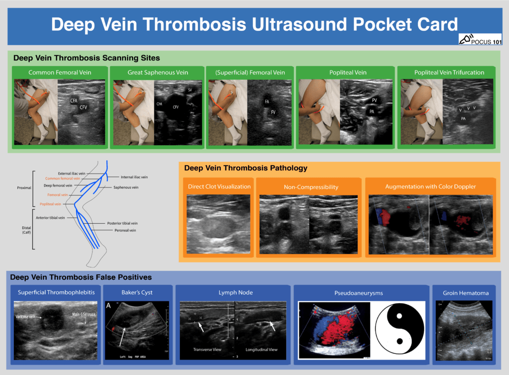
3 These are the most important deep veins to know:
 https://abs.twimg.com/emoji/v2/... draggable="false" alt="1⃣" title="Tastenkappe Ziffer 1" aria-label="Emoji: Tastenkappe Ziffer 1">Common Femoral Vein (CFV)
 https://abs.twimg.com/emoji/v2/... draggable="false" alt="2⃣" title="Tastenkappe Ziffer 2" aria-label="Emoji: Tastenkappe Ziffer 2"> Great Saphenous Vein
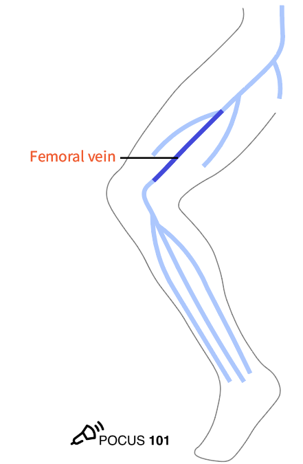
 https://abs.twimg.com/emoji/v2/... draggable="false" alt="3⃣" title="Tastenkappe Ziffer 3" aria-label="Emoji: Tastenkappe Ziffer 3"> Bifurcation of CFV into Deep Femoral Vein and Femoral Vein (aka superficial femoral vein)
 https://abs.twimg.com/emoji/v2/... draggable="false" alt="4⃣" title="Tastenkappe Ziffer 4" aria-label="Emoji: Tastenkappe Ziffer 4"> Popliteal Vein
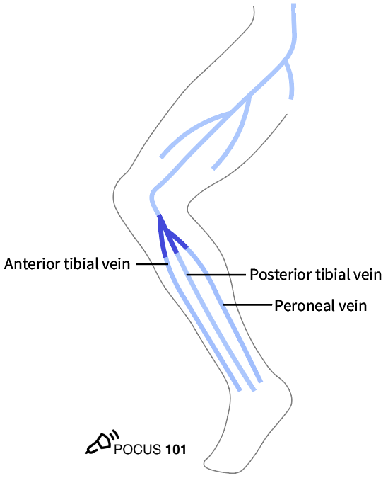
 https://abs.twimg.com/emoji/v2/... draggable="false" alt="5⃣" title="Tastenkappe Ziffer 5" aria-label="Emoji: Tastenkappe Ziffer 5"> Trifurcation of the Popliteal Vein
 https://abs.twimg.com/emoji/v2/... draggable="false" alt="👉" title="Rückhand Zeigefinger nach rechts" aria-label="Emoji: Rückhand Zeigefinger nach rechts">
 https://abs.twimg.com/emoji/v2/... draggable="false" alt="🔗" title="Link Symbol" aria-label="Emoji: Link Symbol"> https://pocus101.com/dvt ">https://pocus101.com/dvt"...
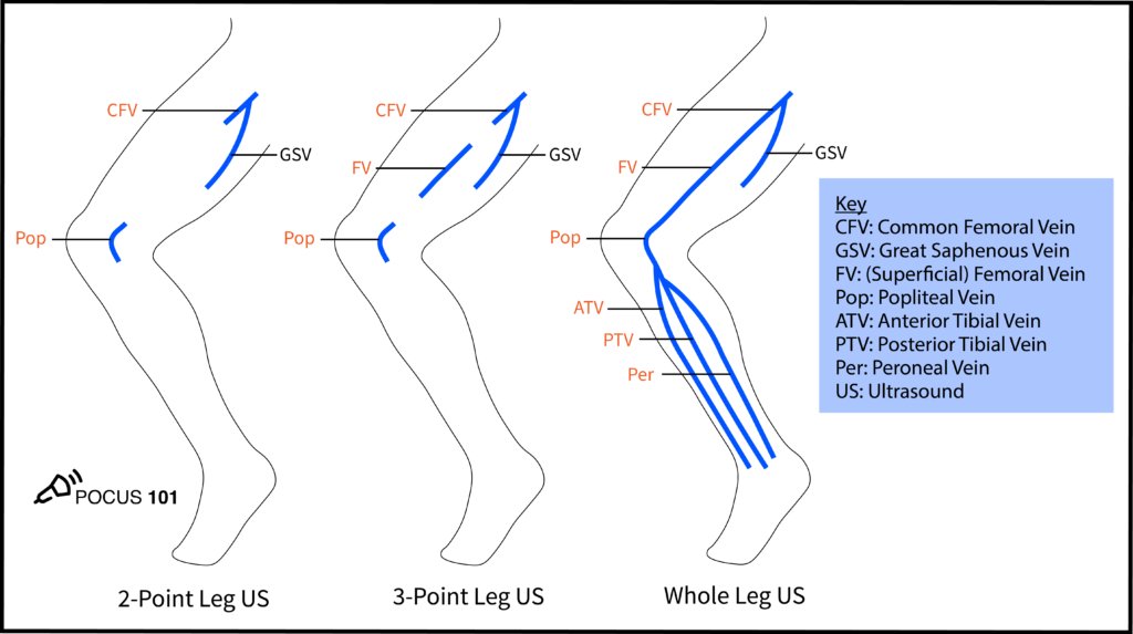
4 We recommend the 3-point protocol
 https://abs.twimg.com/emoji/v2/... draggable="false" alt="👉" title="Rückhand Zeigefinger nach rechts" aria-label="Emoji: Rückhand Zeigefinger nach rechts">
 https://abs.twimg.com/emoji/v2/... draggable="false" alt="🔗" title="Link Symbol" aria-label="Emoji: Link Symbol"> https://pocus101.com/dvt
Compress">https://pocus101.com/dvt"... at:
 https://abs.twimg.com/emoji/v2/... draggable="false" alt="1⃣" title="Tastenkappe Ziffer 1" aria-label="Emoji: Tastenkappe Ziffer 1"> Saphenofemoral Junction
 https://abs.twimg.com/emoji/v2/... draggable="false" alt="2⃣" title="Tastenkappe Ziffer 2" aria-label="Emoji: Tastenkappe Ziffer 2"> Bifurcation of CFV into Deep Femoral Vein and Femoral Vein
 https://abs.twimg.com/emoji/v2/... draggable="false" alt="3⃣" title="Tastenkappe Ziffer 3" aria-label="Emoji: Tastenkappe Ziffer 3"> Popliteal Vein up to the trifurcation
This figure shows the differences b/n protocols
Compress">https://pocus101.com/dvt"... at:
This figure shows the differences b/n protocols
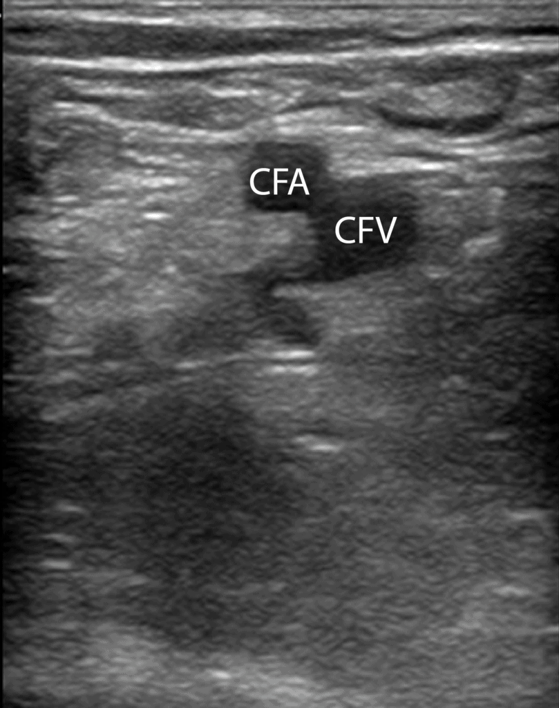
5 Proper Vein Compression: You should apply pressure until the pulsatile artery compresses slightly. If the adjacent vein compresses completely, there is no DVT at that spot.
 https://abs.twimg.com/emoji/v2/... draggable="false" alt="👉" title="Rückhand Zeigefinger nach rechts" aria-label="Emoji: Rückhand Zeigefinger nach rechts">
 https://abs.twimg.com/emoji/v2/... draggable="false" alt="🔗" title="Link Symbol" aria-label="Emoji: Link Symbol"> https://pocus101.com/dvt ">https://pocus101.com/dvt"...
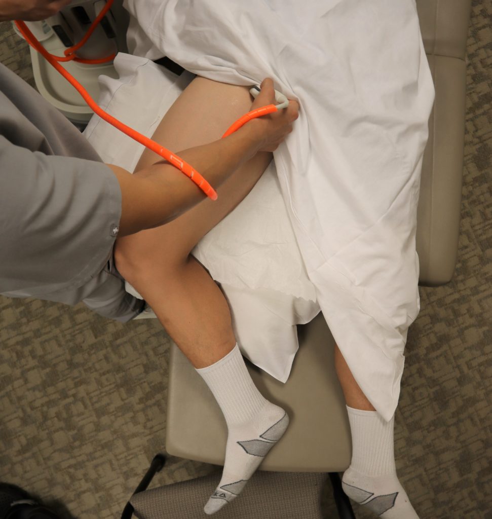
6 Apply gel onto the probe and place it along the inguinal ligament.
Orient the probe perpendicular to the skin with indicator facing the patient’s right to obtain the transverse view of the Common Femoral Vein and Common Femoral Artery. Compress.
 https://abs.twimg.com/emoji/v2/... draggable="false" alt="👉" title="Rückhand Zeigefinger nach rechts" aria-label="Emoji: Rückhand Zeigefinger nach rechts">
 https://abs.twimg.com/emoji/v2/... draggable="false" alt="🔗" title="Link Symbol" aria-label="Emoji: Link Symbol"> https://pocus101.com/dvt ">https://pocus101.com/dvt"...
Orient the probe perpendicular to the skin with indicator facing the patient’s right to obtain the transverse view of the Common Femoral Vein and Common Femoral Artery. Compress.
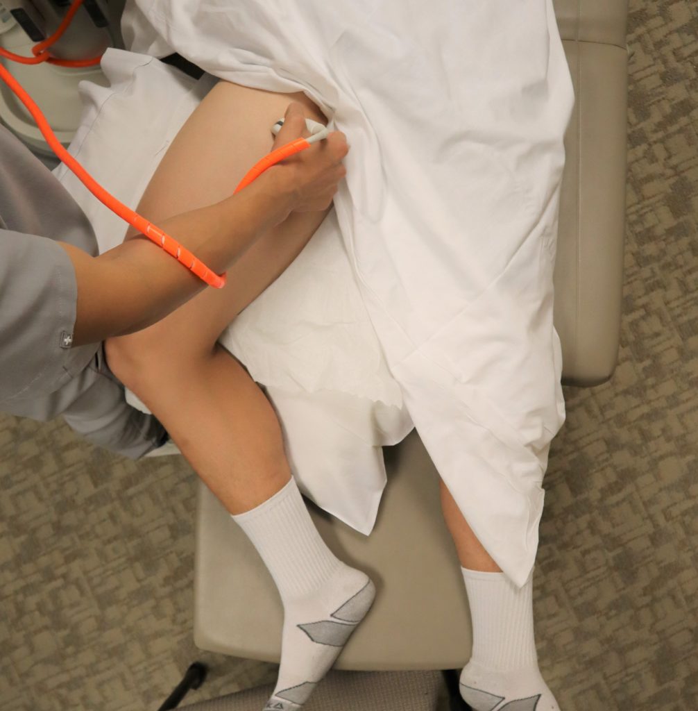
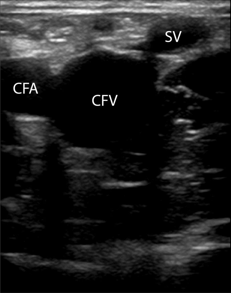
7 Slide the probe 1-2 cm down the patient’s leg to find where the great saphenous vein branches off of the CFV. Apply compression.
 https://abs.twimg.com/emoji/v2/... draggable="false" alt="👉" title="Rückhand Zeigefinger nach rechts" aria-label="Emoji: Rückhand Zeigefinger nach rechts">
 https://abs.twimg.com/emoji/v2/... draggable="false" alt="🔗" title="Link Symbol" aria-label="Emoji: Link Symbol"> https://pocus101.com/dvt ">https://pocus101.com/dvt"...
8 Slide the probe 1-2 cm down the patient’s leg to find where the CFV branches into the deep femoral vein and (superficial) femoral vein. Apply compression.
 https://abs.twimg.com/emoji/v2/... draggable="false" alt="👉" title="Rückhand Zeigefinger nach rechts" aria-label="Emoji: Rückhand Zeigefinger nach rechts">
 https://abs.twimg.com/emoji/v2/... draggable="false" alt="🔗" title="Link Symbol" aria-label="Emoji: Link Symbol"> https://pocus101.com/dvt ">https://pocus101.com/dvt"...
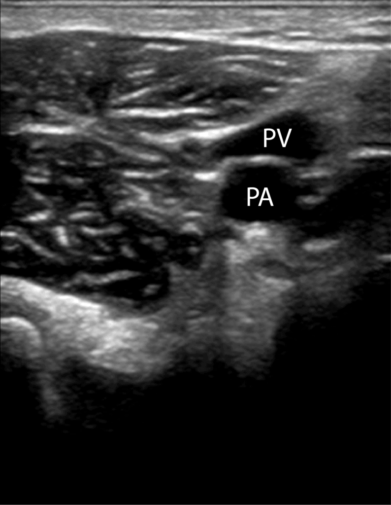
9 Move the probe into the posterior crease of the knee and scan 2 cm above and below to find the popliteal vein. Remember "Pop on Top." Apply compression.
 https://abs.twimg.com/emoji/v2/... draggable="false" alt="👉" title="Rückhand Zeigefinger nach rechts" aria-label="Emoji: Rückhand Zeigefinger nach rechts">
 https://abs.twimg.com/emoji/v2/... draggable="false" alt="🔗" title="Link Symbol" aria-label="Emoji: Link Symbol"> https://pocus101.com/dvt ">https://pocus101.com/dvt"...
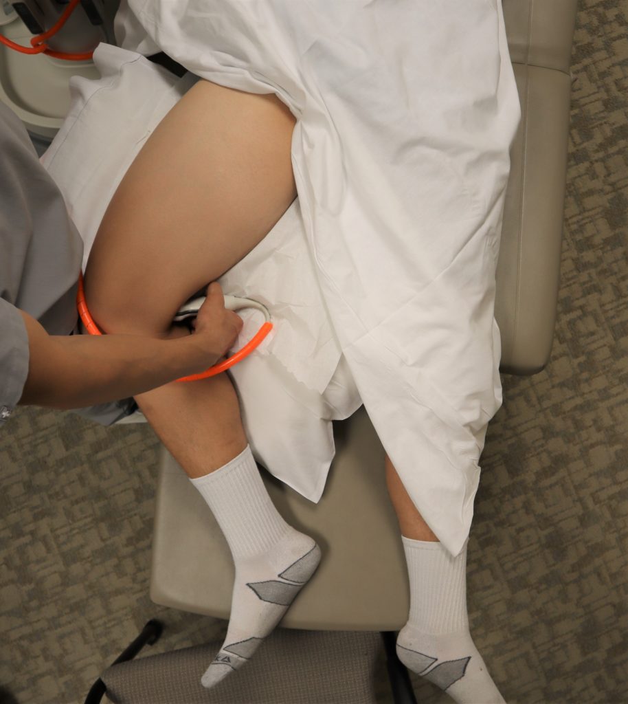
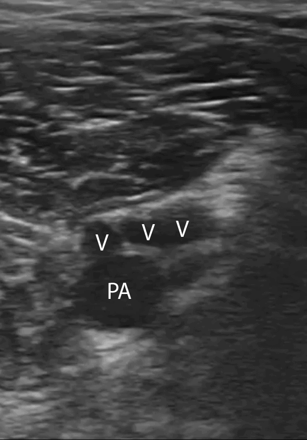
10 Continue to scan slightly more distal from the popliteal vein to find its trifurcation.
 https://abs.twimg.com/emoji/v2/... draggable="false" alt="👉" title="Rückhand Zeigefinger nach rechts" aria-label="Emoji: Rückhand Zeigefinger nach rechts">
 https://abs.twimg.com/emoji/v2/... draggable="false" alt="🔗" title="Link Symbol" aria-label="Emoji: Link Symbol"> https://pocus101.com/dvt ">https://pocus101.com/dvt"...
11 Deep vein thrombosis can be detected in two main ways using point of care ultrasound: direct clot visualization, non-compressibility of the vein. Download the PDF!
 https://abs.twimg.com/emoji/v2/... draggable="false" alt="👉" title="Rückhand Zeigefinger nach rechts" aria-label="Emoji: Rückhand Zeigefinger nach rechts">
 https://abs.twimg.com/emoji/v2/... draggable="false" alt="🔗" title="Link Symbol" aria-label="Emoji: Link Symbol"> https://pocus101.com/dvt ">https://pocus101.com/dvt"...
12 Direct Clot Visualization of echogenic mobile DVT
 https://abs.twimg.com/emoji/v2/... draggable="false" alt="👉" title="Rückhand Zeigefinger nach rechts" aria-label="Emoji: Rückhand Zeigefinger nach rechts">
 https://abs.twimg.com/emoji/v2/... draggable="false" alt="🔗" title="Link Symbol" aria-label="Emoji: Link Symbol"> https://pocus101.com/dvt ">https://pocus101.com/dvt"...
13 Non-compressible vein:
 https://abs.twimg.com/emoji/v2/... draggable="false" alt="👉" title="Rückhand Zeigefinger nach rechts" aria-label="Emoji: Rückhand Zeigefinger nach rechts">
 https://abs.twimg.com/emoji/v2/... draggable="false" alt="🔗" title="Link Symbol" aria-label="Emoji: Link Symbol"> https://pocus101.com/dvt ">https://pocus101.com/dvt"...
14 Color Doppler with Augmentation (squeezing of calf) can be performed but be careful to not dislodge a large or mobile clot!
 https://abs.twimg.com/emoji/v2/... draggable="false" alt="👉" title="Rückhand Zeigefinger nach rechts" aria-label="Emoji: Rückhand Zeigefinger nach rechts">
 https://abs.twimg.com/emoji/v2/... draggable="false" alt="🔗" title="Link Symbol" aria-label="Emoji: Link Symbol"> https://pocus101.com/dvt ">https://pocus101.com/dvt"...
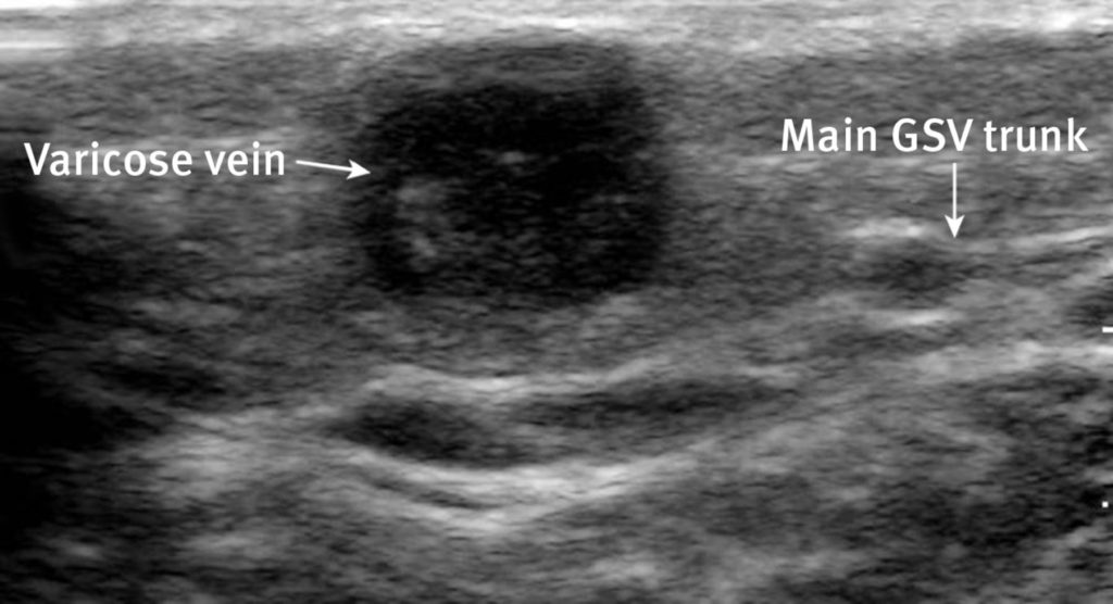
15 Superficial Thrombophlebitis in a varicose vein or small saphenous vein can cause a false positive for DVT.
 https://abs.twimg.com/emoji/v2/... draggable="false" alt="👉" title="Rückhand Zeigefinger nach rechts" aria-label="Emoji: Rückhand Zeigefinger nach rechts">
 https://abs.twimg.com/emoji/v2/... draggable="false" alt="🔗" title="Link Symbol" aria-label="Emoji: Link Symbol"> https://pocus101.com/dvt ">https://pocus101.com/dvt"...
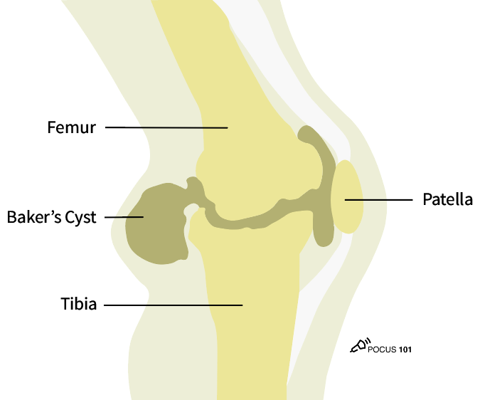
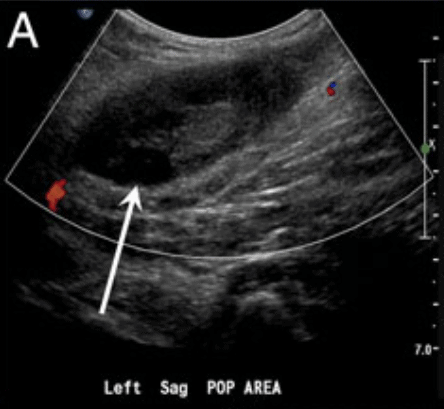
16 A Baker’s cyst is a fluid-filled cyst in the popliteal bursa and can be a false positive.
It appears as a circular anechoic mass with sharply defined borders in both the longitudinal and transverse view. On Color Doppler, there should be no flow.
 https://abs.twimg.com/emoji/v2/... draggable="false" alt="👉" title="Rückhand Zeigefinger nach rechts" aria-label="Emoji: Rückhand Zeigefinger nach rechts">
 https://abs.twimg.com/emoji/v2/... draggable="false" alt="🔗" title="Link Symbol" aria-label="Emoji: Link Symbol"> https://pocus101.com/dvt ">https://pocus101.com/dvt"...
It appears as a circular anechoic mass with sharply defined borders in both the longitudinal and transverse view. On Color Doppler, there should be no flow.
17 On ultrasound, a lymph node can look similar to a clot because it is a hypoechoic oval structure with a hyperechoic center. However, in the longitudinal view, a lymph node is circumscribed and not tubular in structure like a vein.
 https://abs.twimg.com/emoji/v2/... draggable="false" alt="👉" title="Rückhand Zeigefinger nach rechts" aria-label="Emoji: Rückhand Zeigefinger nach rechts">
 https://abs.twimg.com/emoji/v2/... draggable="false" alt="🔗" title="Link Symbol" aria-label="Emoji: Link Symbol"> https://pocus101.com/dvt ">https://pocus101.com/dvt"...
18 On ultrasound, pseudoaneurysms present as anechoic or hypoechoic images. With Color Doppler, you can see a “Yin-Yang Sign” from the circular motion of blood inside the pseudoaneurysm cavity. Don& #39;t confuse this for a non-compressible vein.
 https://abs.twimg.com/emoji/v2/... draggable="false" alt="👉" title="Rückhand Zeigefinger nach rechts" aria-label="Emoji: Rückhand Zeigefinger nach rechts">
 https://abs.twimg.com/emoji/v2/... draggable="false" alt="🔗" title="Link Symbol" aria-label="Emoji: Link Symbol"> https://pocus101.com/dvt ">https://pocus101.com/dvt"...
19 On ultrasound, a groin hematoma will be hypoechoic with some anechoic areas scattered throughout. If you scan the hematoma in the longitudinal view, it becomes obvious that the object is circumscribed and not tubular like a vein or artery.
 https://abs.twimg.com/emoji/v2/... draggable="false" alt="👉" title="Rückhand Zeigefinger nach rechts" aria-label="Emoji: Rückhand Zeigefinger nach rechts">
 https://abs.twimg.com/emoji/v2/... draggable="false" alt="🔗" title="Link Symbol" aria-label="Emoji: Link Symbol"> https://pocus101.com/dvt ">https://pocus101.com/dvt"...
20 Want to learn even MORE about DVT ultrasound? Check out our newly released #POCUS Board Review Book with an exclusive 20% discount code!
 https://abs.twimg.com/emoji/v2/... draggable="false" alt="👉" title="Rückhand Zeigefinger nach rechts" aria-label="Emoji: Rückhand Zeigefinger nach rechts">
 https://abs.twimg.com/emoji/v2/... draggable="false" alt="🔗" title="Link Symbol" aria-label="Emoji: Link Symbol"> https://pocus101.com/RevBook ">https://pocus101.com/RevBook&q...

 Read on Twitter
 + https://abs.twimg.com/emoji/v2/... draggable="false" alt="🫁" title="Lungs" aria-label="Emoji: Lungs">! #POCUShttps://abs.twimg.com/emoji/v2/... draggable="false" alt="1⃣" title="Tastenkappe Ziffer 1" aria-label="Emoji: Tastenkappe Ziffer 1">Learn how to Easily do DVT Ultrasoundhttps://abs.twimg.com/emoji/v2/... draggable="false" alt="2⃣" title="Tastenkappe Ziffer 2" aria-label="Emoji: Tastenkappe Ziffer 2">Learn how to use Compression Properlyhttps://abs.twimg.com/emoji/v2/... draggable="false" alt="3⃣" title="Tastenkappe Ziffer 3" aria-label="Emoji: Tastenkappe Ziffer 3">Recognize False Positives for DVThttps://abs.twimg.com/emoji/v2/... draggable="false" alt="✅" title="Fettes weißes Häkchen" aria-label="Emoji: Fettes weißes Häkchen">New Blog Post https://abs.twimg.com/emoji/v2/... draggable="false" alt="👉" title="Rückhand Zeigefinger nach rechts" aria-label="Emoji: Rückhand Zeigefinger nach rechts">https://abs.twimg.com/emoji/v2/... draggable="false" alt="🔗" title="Link Symbol" aria-label="Emoji: Link Symbol"> https://pocus101.com/dvt"... href="https://twtext.com//hashtag/medtweetorial"> #medtweetorialhttps://abs.twimg.com/emoji/v2/... draggable="false" alt="👇" title="Rückhand Zeigefinger nach unten" aria-label="Emoji: Rückhand Zeigefinger nach unten">(1/20)" title="Catch those DVTs Early... BEFORE they get to the https://abs.twimg.com/emoji/v2/... draggable="false" alt="💔" title="Gebrochenes Herz" aria-label="Emoji: Gebrochenes Herz"> + https://abs.twimg.com/emoji/v2/... draggable="false" alt="🫁" title="Lungs" aria-label="Emoji: Lungs">! #POCUShttps://abs.twimg.com/emoji/v2/... draggable="false" alt="1⃣" title="Tastenkappe Ziffer 1" aria-label="Emoji: Tastenkappe Ziffer 1">Learn how to Easily do DVT Ultrasoundhttps://abs.twimg.com/emoji/v2/... draggable="false" alt="2⃣" title="Tastenkappe Ziffer 2" aria-label="Emoji: Tastenkappe Ziffer 2">Learn how to use Compression Properlyhttps://abs.twimg.com/emoji/v2/... draggable="false" alt="3⃣" title="Tastenkappe Ziffer 3" aria-label="Emoji: Tastenkappe Ziffer 3">Recognize False Positives for DVThttps://abs.twimg.com/emoji/v2/... draggable="false" alt="✅" title="Fettes weißes Häkchen" aria-label="Emoji: Fettes weißes Häkchen">New Blog Post https://abs.twimg.com/emoji/v2/... draggable="false" alt="👉" title="Rückhand Zeigefinger nach rechts" aria-label="Emoji: Rückhand Zeigefinger nach rechts">https://abs.twimg.com/emoji/v2/... draggable="false" alt="🔗" title="Link Symbol" aria-label="Emoji: Link Symbol"> https://pocus101.com/dvt"... href="https://twtext.com//hashtag/medtweetorial"> #medtweetorialhttps://abs.twimg.com/emoji/v2/... draggable="false" alt="👇" title="Rückhand Zeigefinger nach unten" aria-label="Emoji: Rückhand Zeigefinger nach unten">(1/20)">
 + https://abs.twimg.com/emoji/v2/... draggable="false" alt="🫁" title="Lungs" aria-label="Emoji: Lungs">! #POCUShttps://abs.twimg.com/emoji/v2/... draggable="false" alt="1⃣" title="Tastenkappe Ziffer 1" aria-label="Emoji: Tastenkappe Ziffer 1">Learn how to Easily do DVT Ultrasoundhttps://abs.twimg.com/emoji/v2/... draggable="false" alt="2⃣" title="Tastenkappe Ziffer 2" aria-label="Emoji: Tastenkappe Ziffer 2">Learn how to use Compression Properlyhttps://abs.twimg.com/emoji/v2/... draggable="false" alt="3⃣" title="Tastenkappe Ziffer 3" aria-label="Emoji: Tastenkappe Ziffer 3">Recognize False Positives for DVThttps://abs.twimg.com/emoji/v2/... draggable="false" alt="✅" title="Fettes weißes Häkchen" aria-label="Emoji: Fettes weißes Häkchen">New Blog Post https://abs.twimg.com/emoji/v2/... draggable="false" alt="👉" title="Rückhand Zeigefinger nach rechts" aria-label="Emoji: Rückhand Zeigefinger nach rechts">https://abs.twimg.com/emoji/v2/... draggable="false" alt="🔗" title="Link Symbol" aria-label="Emoji: Link Symbol"> https://pocus101.com/dvt"... href="https://twtext.com//hashtag/medtweetorial"> #medtweetorialhttps://abs.twimg.com/emoji/v2/... draggable="false" alt="👇" title="Rückhand Zeigefinger nach unten" aria-label="Emoji: Rückhand Zeigefinger nach unten">(1/20)" title="Catch those DVTs Early... BEFORE they get to the https://abs.twimg.com/emoji/v2/... draggable="false" alt="💔" title="Gebrochenes Herz" aria-label="Emoji: Gebrochenes Herz"> + https://abs.twimg.com/emoji/v2/... draggable="false" alt="🫁" title="Lungs" aria-label="Emoji: Lungs">! #POCUShttps://abs.twimg.com/emoji/v2/... draggable="false" alt="1⃣" title="Tastenkappe Ziffer 1" aria-label="Emoji: Tastenkappe Ziffer 1">Learn how to Easily do DVT Ultrasoundhttps://abs.twimg.com/emoji/v2/... draggable="false" alt="2⃣" title="Tastenkappe Ziffer 2" aria-label="Emoji: Tastenkappe Ziffer 2">Learn how to use Compression Properlyhttps://abs.twimg.com/emoji/v2/... draggable="false" alt="3⃣" title="Tastenkappe Ziffer 3" aria-label="Emoji: Tastenkappe Ziffer 3">Recognize False Positives for DVThttps://abs.twimg.com/emoji/v2/... draggable="false" alt="✅" title="Fettes weißes Häkchen" aria-label="Emoji: Fettes weißes Häkchen">New Blog Post https://abs.twimg.com/emoji/v2/... draggable="false" alt="👉" title="Rückhand Zeigefinger nach rechts" aria-label="Emoji: Rückhand Zeigefinger nach rechts">https://abs.twimg.com/emoji/v2/... draggable="false" alt="🔗" title="Link Symbol" aria-label="Emoji: Link Symbol"> https://pocus101.com/dvt"... href="https://twtext.com//hashtag/medtweetorial"> #medtweetorialhttps://abs.twimg.com/emoji/v2/... draggable="false" alt="👇" title="Rückhand Zeigefinger nach unten" aria-label="Emoji: Rückhand Zeigefinger nach unten">(1/20)">
 + https://abs.twimg.com/emoji/v2/... draggable="false" alt="🫁" title="Lungs" aria-label="Emoji: Lungs">! #POCUShttps://abs.twimg.com/emoji/v2/... draggable="false" alt="1⃣" title="Tastenkappe Ziffer 1" aria-label="Emoji: Tastenkappe Ziffer 1">Learn how to Easily do DVT Ultrasoundhttps://abs.twimg.com/emoji/v2/... draggable="false" alt="2⃣" title="Tastenkappe Ziffer 2" aria-label="Emoji: Tastenkappe Ziffer 2">Learn how to use Compression Properlyhttps://abs.twimg.com/emoji/v2/... draggable="false" alt="3⃣" title="Tastenkappe Ziffer 3" aria-label="Emoji: Tastenkappe Ziffer 3">Recognize False Positives for DVThttps://abs.twimg.com/emoji/v2/... draggable="false" alt="✅" title="Fettes weißes Häkchen" aria-label="Emoji: Fettes weißes Häkchen">New Blog Post https://abs.twimg.com/emoji/v2/... draggable="false" alt="👉" title="Rückhand Zeigefinger nach rechts" aria-label="Emoji: Rückhand Zeigefinger nach rechts">https://abs.twimg.com/emoji/v2/... draggable="false" alt="🔗" title="Link Symbol" aria-label="Emoji: Link Symbol"> https://pocus101.com/dvt"... href="https://twtext.com//hashtag/medtweetorial"> #medtweetorialhttps://abs.twimg.com/emoji/v2/... draggable="false" alt="👇" title="Rückhand Zeigefinger nach unten" aria-label="Emoji: Rückhand Zeigefinger nach unten">(1/20)" title="Catch those DVTs Early... BEFORE they get to the https://abs.twimg.com/emoji/v2/... draggable="false" alt="💔" title="Gebrochenes Herz" aria-label="Emoji: Gebrochenes Herz"> + https://abs.twimg.com/emoji/v2/... draggable="false" alt="🫁" title="Lungs" aria-label="Emoji: Lungs">! #POCUShttps://abs.twimg.com/emoji/v2/... draggable="false" alt="1⃣" title="Tastenkappe Ziffer 1" aria-label="Emoji: Tastenkappe Ziffer 1">Learn how to Easily do DVT Ultrasoundhttps://abs.twimg.com/emoji/v2/... draggable="false" alt="2⃣" title="Tastenkappe Ziffer 2" aria-label="Emoji: Tastenkappe Ziffer 2">Learn how to use Compression Properlyhttps://abs.twimg.com/emoji/v2/... draggable="false" alt="3⃣" title="Tastenkappe Ziffer 3" aria-label="Emoji: Tastenkappe Ziffer 3">Recognize False Positives for DVThttps://abs.twimg.com/emoji/v2/... draggable="false" alt="✅" title="Fettes weißes Häkchen" aria-label="Emoji: Fettes weißes Häkchen">New Blog Post https://abs.twimg.com/emoji/v2/... draggable="false" alt="👉" title="Rückhand Zeigefinger nach rechts" aria-label="Emoji: Rückhand Zeigefinger nach rechts">https://abs.twimg.com/emoji/v2/... draggable="false" alt="🔗" title="Link Symbol" aria-label="Emoji: Link Symbol"> https://pocus101.com/dvt"... href="https://twtext.com//hashtag/medtweetorial"> #medtweetorialhttps://abs.twimg.com/emoji/v2/... draggable="false" alt="👇" title="Rückhand Zeigefinger nach unten" aria-label="Emoji: Rückhand Zeigefinger nach unten">(1/20)">
 https://abs.twimg.com/emoji/v2/... draggable="false" alt="🔗" title="Link Symbol" aria-label="Emoji: Link Symbol"> https://pocus101.com/dvt"..." title="2 Download the FREE DVT Ultrasound PDF Pocket Card!https://abs.twimg.com/emoji/v2/... draggable="false" alt="👉" title="Rückhand Zeigefinger nach rechts" aria-label="Emoji: Rückhand Zeigefinger nach rechts">https://abs.twimg.com/emoji/v2/... draggable="false" alt="🔗" title="Link Symbol" aria-label="Emoji: Link Symbol"> https://pocus101.com/dvt"..." class="img-responsive" style="max-width:100%;"/>
 Common Femoral Vein (CFV)https://abs.twimg.com/emoji/v2/... draggable="false" alt="2⃣" title="Tastenkappe Ziffer 2" aria-label="Emoji: Tastenkappe Ziffer 2"> Great Saphenous Veinhttps://abs.twimg.com/emoji/v2/... draggable="false" alt="3⃣" title="Tastenkappe Ziffer 3" aria-label="Emoji: Tastenkappe Ziffer 3"> Bifurcation of CFV into Deep Femoral Vein and Femoral Vein (aka superficial femoral vein)https://abs.twimg.com/emoji/v2/... draggable="false" alt="4⃣" title="Tastenkappe Ziffer 4" aria-label="Emoji: Tastenkappe Ziffer 4"> Popliteal Veinhttps://abs.twimg.com/emoji/v2/... draggable="false" alt="5⃣" title="Tastenkappe Ziffer 5" aria-label="Emoji: Tastenkappe Ziffer 5"> Trifurcation of the Popliteal Veinhttps://abs.twimg.com/emoji/v2/... draggable="false" alt="👉" title="Rückhand Zeigefinger nach rechts" aria-label="Emoji: Rückhand Zeigefinger nach rechts">https://abs.twimg.com/emoji/v2/... draggable="false" alt="🔗" title="Link Symbol" aria-label="Emoji: Link Symbol"> https://pocus101.com/dvt"..." title="3 These are the most important deep veins to know:https://abs.twimg.com/emoji/v2/... draggable="false" alt="1⃣" title="Tastenkappe Ziffer 1" aria-label="Emoji: Tastenkappe Ziffer 1">Common Femoral Vein (CFV)https://abs.twimg.com/emoji/v2/... draggable="false" alt="2⃣" title="Tastenkappe Ziffer 2" aria-label="Emoji: Tastenkappe Ziffer 2"> Great Saphenous Veinhttps://abs.twimg.com/emoji/v2/... draggable="false" alt="3⃣" title="Tastenkappe Ziffer 3" aria-label="Emoji: Tastenkappe Ziffer 3"> Bifurcation of CFV into Deep Femoral Vein and Femoral Vein (aka superficial femoral vein)https://abs.twimg.com/emoji/v2/... draggable="false" alt="4⃣" title="Tastenkappe Ziffer 4" aria-label="Emoji: Tastenkappe Ziffer 4"> Popliteal Veinhttps://abs.twimg.com/emoji/v2/... draggable="false" alt="5⃣" title="Tastenkappe Ziffer 5" aria-label="Emoji: Tastenkappe Ziffer 5"> Trifurcation of the Popliteal Veinhttps://abs.twimg.com/emoji/v2/... draggable="false" alt="👉" title="Rückhand Zeigefinger nach rechts" aria-label="Emoji: Rückhand Zeigefinger nach rechts">https://abs.twimg.com/emoji/v2/... draggable="false" alt="🔗" title="Link Symbol" aria-label="Emoji: Link Symbol"> https://pocus101.com/dvt"..." class="img-responsive" style="max-width:100%;"/>
 https://abs.twimg.com/emoji/v2/... draggable="false" alt="🔗" title="Link Symbol" aria-label="Emoji: Link Symbol"> https://pocus101.com/dvt"... at:https://abs.twimg.com/emoji/v2/... draggable="false" alt="1⃣" title="Tastenkappe Ziffer 1" aria-label="Emoji: Tastenkappe Ziffer 1"> Saphenofemoral Junctionhttps://abs.twimg.com/emoji/v2/... draggable="false" alt="2⃣" title="Tastenkappe Ziffer 2" aria-label="Emoji: Tastenkappe Ziffer 2"> Bifurcation of CFV into Deep Femoral Vein and Femoral Veinhttps://abs.twimg.com/emoji/v2/... draggable="false" alt="3⃣" title="Tastenkappe Ziffer 3" aria-label="Emoji: Tastenkappe Ziffer 3"> Popliteal Vein up to the trifurcationThis figure shows the differences b/n protocols" title="4 We recommend the 3-point protocolhttps://abs.twimg.com/emoji/v2/... draggable="false" alt="👉" title="Rückhand Zeigefinger nach rechts" aria-label="Emoji: Rückhand Zeigefinger nach rechts">https://abs.twimg.com/emoji/v2/... draggable="false" alt="🔗" title="Link Symbol" aria-label="Emoji: Link Symbol"> https://pocus101.com/dvt"... at:https://abs.twimg.com/emoji/v2/... draggable="false" alt="1⃣" title="Tastenkappe Ziffer 1" aria-label="Emoji: Tastenkappe Ziffer 1"> Saphenofemoral Junctionhttps://abs.twimg.com/emoji/v2/... draggable="false" alt="2⃣" title="Tastenkappe Ziffer 2" aria-label="Emoji: Tastenkappe Ziffer 2"> Bifurcation of CFV into Deep Femoral Vein and Femoral Veinhttps://abs.twimg.com/emoji/v2/... draggable="false" alt="3⃣" title="Tastenkappe Ziffer 3" aria-label="Emoji: Tastenkappe Ziffer 3"> Popliteal Vein up to the trifurcationThis figure shows the differences b/n protocols" class="img-responsive" style="max-width:100%;"/>
 https://abs.twimg.com/emoji/v2/... draggable="false" alt="🔗" title="Link Symbol" aria-label="Emoji: Link Symbol"> https://pocus101.com/dvt"..." title="6 Apply gel onto the probe and place it along the inguinal ligament.Orient the probe perpendicular to the skin with indicator facing the patient’s right to obtain the transverse view of the Common Femoral Vein and Common Femoral Artery. Compress.https://abs.twimg.com/emoji/v2/... draggable="false" alt="👉" title="Rückhand Zeigefinger nach rechts" aria-label="Emoji: Rückhand Zeigefinger nach rechts">https://abs.twimg.com/emoji/v2/... draggable="false" alt="🔗" title="Link Symbol" aria-label="Emoji: Link Symbol"> https://pocus101.com/dvt"...">
 https://abs.twimg.com/emoji/v2/... draggable="false" alt="🔗" title="Link Symbol" aria-label="Emoji: Link Symbol"> https://pocus101.com/dvt"..." title="6 Apply gel onto the probe and place it along the inguinal ligament.Orient the probe perpendicular to the skin with indicator facing the patient’s right to obtain the transverse view of the Common Femoral Vein and Common Femoral Artery. Compress.https://abs.twimg.com/emoji/v2/... draggable="false" alt="👉" title="Rückhand Zeigefinger nach rechts" aria-label="Emoji: Rückhand Zeigefinger nach rechts">https://abs.twimg.com/emoji/v2/... draggable="false" alt="🔗" title="Link Symbol" aria-label="Emoji: Link Symbol"> https://pocus101.com/dvt"...">
 https://abs.twimg.com/emoji/v2/... draggable="false" alt="🔗" title="Link Symbol" aria-label="Emoji: Link Symbol"> https://pocus101.com/dvt"..." title="6 Apply gel onto the probe and place it along the inguinal ligament.Orient the probe perpendicular to the skin with indicator facing the patient’s right to obtain the transverse view of the Common Femoral Vein and Common Femoral Artery. Compress.https://abs.twimg.com/emoji/v2/... draggable="false" alt="👉" title="Rückhand Zeigefinger nach rechts" aria-label="Emoji: Rückhand Zeigefinger nach rechts">https://abs.twimg.com/emoji/v2/... draggable="false" alt="🔗" title="Link Symbol" aria-label="Emoji: Link Symbol"> https://pocus101.com/dvt"...">
 https://abs.twimg.com/emoji/v2/... draggable="false" alt="🔗" title="Link Symbol" aria-label="Emoji: Link Symbol"> https://pocus101.com/dvt"..." title="7 Slide the probe 1-2 cm down the patient’s leg to find where the great saphenous vein branches off of the CFV. Apply compression.https://abs.twimg.com/emoji/v2/... draggable="false" alt="👉" title="Rückhand Zeigefinger nach rechts" aria-label="Emoji: Rückhand Zeigefinger nach rechts">https://abs.twimg.com/emoji/v2/... draggable="false" alt="🔗" title="Link Symbol" aria-label="Emoji: Link Symbol"> https://pocus101.com/dvt"...">
 https://abs.twimg.com/emoji/v2/... draggable="false" alt="🔗" title="Link Symbol" aria-label="Emoji: Link Symbol"> https://pocus101.com/dvt"..." title="7 Slide the probe 1-2 cm down the patient’s leg to find where the great saphenous vein branches off of the CFV. Apply compression.https://abs.twimg.com/emoji/v2/... draggable="false" alt="👉" title="Rückhand Zeigefinger nach rechts" aria-label="Emoji: Rückhand Zeigefinger nach rechts">https://abs.twimg.com/emoji/v2/... draggable="false" alt="🔗" title="Link Symbol" aria-label="Emoji: Link Symbol"> https://pocus101.com/dvt"...">
 https://abs.twimg.com/emoji/v2/... draggable="false" alt="🔗" title="Link Symbol" aria-label="Emoji: Link Symbol"> https://pocus101.com/dvt"..." title="7 Slide the probe 1-2 cm down the patient’s leg to find where the great saphenous vein branches off of the CFV. Apply compression.https://abs.twimg.com/emoji/v2/... draggable="false" alt="👉" title="Rückhand Zeigefinger nach rechts" aria-label="Emoji: Rückhand Zeigefinger nach rechts">https://abs.twimg.com/emoji/v2/... draggable="false" alt="🔗" title="Link Symbol" aria-label="Emoji: Link Symbol"> https://pocus101.com/dvt"...">
 https://abs.twimg.com/emoji/v2/... draggable="false" alt="🔗" title="Link Symbol" aria-label="Emoji: Link Symbol"> https://pocus101.com/dvt"..." title="8 Slide the probe 1-2 cm down the patient’s leg to find where the CFV branches into the deep femoral vein and (superficial) femoral vein. Apply compression.https://abs.twimg.com/emoji/v2/... draggable="false" alt="👉" title="Rückhand Zeigefinger nach rechts" aria-label="Emoji: Rückhand Zeigefinger nach rechts">https://abs.twimg.com/emoji/v2/... draggable="false" alt="🔗" title="Link Symbol" aria-label="Emoji: Link Symbol"> https://pocus101.com/dvt"...">
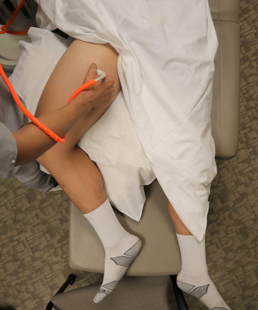 https://abs.twimg.com/emoji/v2/... draggable="false" alt="🔗" title="Link Symbol" aria-label="Emoji: Link Symbol"> https://pocus101.com/dvt"..." title="8 Slide the probe 1-2 cm down the patient’s leg to find where the CFV branches into the deep femoral vein and (superficial) femoral vein. Apply compression.https://abs.twimg.com/emoji/v2/... draggable="false" alt="👉" title="Rückhand Zeigefinger nach rechts" aria-label="Emoji: Rückhand Zeigefinger nach rechts">https://abs.twimg.com/emoji/v2/... draggable="false" alt="🔗" title="Link Symbol" aria-label="Emoji: Link Symbol"> https://pocus101.com/dvt"...">
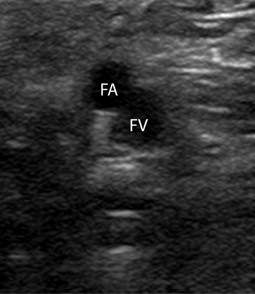 https://abs.twimg.com/emoji/v2/... draggable="false" alt="🔗" title="Link Symbol" aria-label="Emoji: Link Symbol"> https://pocus101.com/dvt"..." title="8 Slide the probe 1-2 cm down the patient’s leg to find where the CFV branches into the deep femoral vein and (superficial) femoral vein. Apply compression.https://abs.twimg.com/emoji/v2/... draggable="false" alt="👉" title="Rückhand Zeigefinger nach rechts" aria-label="Emoji: Rückhand Zeigefinger nach rechts">https://abs.twimg.com/emoji/v2/... draggable="false" alt="🔗" title="Link Symbol" aria-label="Emoji: Link Symbol"> https://pocus101.com/dvt"...">
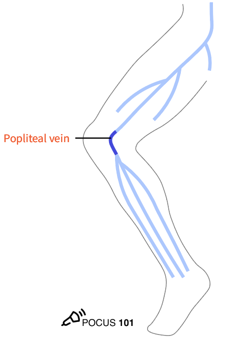 https://abs.twimg.com/emoji/v2/... draggable="false" alt="🔗" title="Link Symbol" aria-label="Emoji: Link Symbol"> https://pocus101.com/dvt"..." title="9 Move the probe into the posterior crease of the knee and scan 2 cm above and below to find the popliteal vein. Remember "Pop on Top." Apply compression.https://abs.twimg.com/emoji/v2/... draggable="false" alt="👉" title="Rückhand Zeigefinger nach rechts" aria-label="Emoji: Rückhand Zeigefinger nach rechts">https://abs.twimg.com/emoji/v2/... draggable="false" alt="🔗" title="Link Symbol" aria-label="Emoji: Link Symbol"> https://pocus101.com/dvt"...">
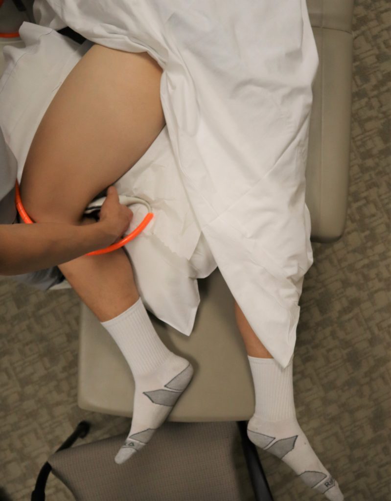 https://abs.twimg.com/emoji/v2/... draggable="false" alt="🔗" title="Link Symbol" aria-label="Emoji: Link Symbol"> https://pocus101.com/dvt"..." title="9 Move the probe into the posterior crease of the knee and scan 2 cm above and below to find the popliteal vein. Remember "Pop on Top." Apply compression.https://abs.twimg.com/emoji/v2/... draggable="false" alt="👉" title="Rückhand Zeigefinger nach rechts" aria-label="Emoji: Rückhand Zeigefinger nach rechts">https://abs.twimg.com/emoji/v2/... draggable="false" alt="🔗" title="Link Symbol" aria-label="Emoji: Link Symbol"> https://pocus101.com/dvt"...">
 https://abs.twimg.com/emoji/v2/... draggable="false" alt="🔗" title="Link Symbol" aria-label="Emoji: Link Symbol"> https://pocus101.com/dvt"..." title="9 Move the probe into the posterior crease of the knee and scan 2 cm above and below to find the popliteal vein. Remember "Pop on Top." Apply compression.https://abs.twimg.com/emoji/v2/... draggable="false" alt="👉" title="Rückhand Zeigefinger nach rechts" aria-label="Emoji: Rückhand Zeigefinger nach rechts">https://abs.twimg.com/emoji/v2/... draggable="false" alt="🔗" title="Link Symbol" aria-label="Emoji: Link Symbol"> https://pocus101.com/dvt"...">
 https://abs.twimg.com/emoji/v2/... draggable="false" alt="🔗" title="Link Symbol" aria-label="Emoji: Link Symbol"> https://pocus101.com/dvt"..." title="10 Continue to scan slightly more distal from the popliteal vein to find its trifurcation.https://abs.twimg.com/emoji/v2/... draggable="false" alt="👉" title="Rückhand Zeigefinger nach rechts" aria-label="Emoji: Rückhand Zeigefinger nach rechts">https://abs.twimg.com/emoji/v2/... draggable="false" alt="🔗" title="Link Symbol" aria-label="Emoji: Link Symbol"> https://pocus101.com/dvt"...">
 https://abs.twimg.com/emoji/v2/... draggable="false" alt="🔗" title="Link Symbol" aria-label="Emoji: Link Symbol"> https://pocus101.com/dvt"..." title="10 Continue to scan slightly more distal from the popliteal vein to find its trifurcation.https://abs.twimg.com/emoji/v2/... draggable="false" alt="👉" title="Rückhand Zeigefinger nach rechts" aria-label="Emoji: Rückhand Zeigefinger nach rechts">https://abs.twimg.com/emoji/v2/... draggable="false" alt="🔗" title="Link Symbol" aria-label="Emoji: Link Symbol"> https://pocus101.com/dvt"...">
 https://abs.twimg.com/emoji/v2/... draggable="false" alt="🔗" title="Link Symbol" aria-label="Emoji: Link Symbol"> https://pocus101.com/dvt"..." title="10 Continue to scan slightly more distal from the popliteal vein to find its trifurcation.https://abs.twimg.com/emoji/v2/... draggable="false" alt="👉" title="Rückhand Zeigefinger nach rechts" aria-label="Emoji: Rückhand Zeigefinger nach rechts">https://abs.twimg.com/emoji/v2/... draggable="false" alt="🔗" title="Link Symbol" aria-label="Emoji: Link Symbol"> https://pocus101.com/dvt"...">
 https://abs.twimg.com/emoji/v2/... draggable="false" alt="🔗" title="Link Symbol" aria-label="Emoji: Link Symbol"> https://pocus101.com/dvt"..." title="11 Deep vein thrombosis can be detected in two main ways using point of care ultrasound: direct clot visualization, non-compressibility of the vein. Download the PDF!https://abs.twimg.com/emoji/v2/... draggable="false" alt="👉" title="Rückhand Zeigefinger nach rechts" aria-label="Emoji: Rückhand Zeigefinger nach rechts">https://abs.twimg.com/emoji/v2/... draggable="false" alt="🔗" title="Link Symbol" aria-label="Emoji: Link Symbol"> https://pocus101.com/dvt"..." class="img-responsive" style="max-width:100%;"/>
 https://abs.twimg.com/emoji/v2/... draggable="false" alt="🔗" title="Link Symbol" aria-label="Emoji: Link Symbol"> https://pocus101.com/dvt"..." title="15 Superficial Thrombophlebitis in a varicose vein or small saphenous vein can cause a false positive for DVT. https://abs.twimg.com/emoji/v2/... draggable="false" alt="👉" title="Rückhand Zeigefinger nach rechts" aria-label="Emoji: Rückhand Zeigefinger nach rechts">https://abs.twimg.com/emoji/v2/... draggable="false" alt="🔗" title="Link Symbol" aria-label="Emoji: Link Symbol"> https://pocus101.com/dvt"..." class="img-responsive" style="max-width:100%;"/>
 https://abs.twimg.com/emoji/v2/... draggable="false" alt="🔗" title="Link Symbol" aria-label="Emoji: Link Symbol"> https://pocus101.com/dvt"..." title="16 A Baker’s cyst is a fluid-filled cyst in the popliteal bursa and can be a false positive.It appears as a circular anechoic mass with sharply defined borders in both the longitudinal and transverse view. On Color Doppler, there should be no flow.https://abs.twimg.com/emoji/v2/... draggable="false" alt="👉" title="Rückhand Zeigefinger nach rechts" aria-label="Emoji: Rückhand Zeigefinger nach rechts">https://abs.twimg.com/emoji/v2/... draggable="false" alt="🔗" title="Link Symbol" aria-label="Emoji: Link Symbol"> https://pocus101.com/dvt"...">
 https://abs.twimg.com/emoji/v2/... draggable="false" alt="🔗" title="Link Symbol" aria-label="Emoji: Link Symbol"> https://pocus101.com/dvt"..." title="16 A Baker’s cyst is a fluid-filled cyst in the popliteal bursa and can be a false positive.It appears as a circular anechoic mass with sharply defined borders in both the longitudinal and transverse view. On Color Doppler, there should be no flow.https://abs.twimg.com/emoji/v2/... draggable="false" alt="👉" title="Rückhand Zeigefinger nach rechts" aria-label="Emoji: Rückhand Zeigefinger nach rechts">https://abs.twimg.com/emoji/v2/... draggable="false" alt="🔗" title="Link Symbol" aria-label="Emoji: Link Symbol"> https://pocus101.com/dvt"...">
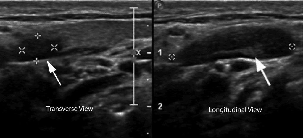 https://abs.twimg.com/emoji/v2/... draggable="false" alt="🔗" title="Link Symbol" aria-label="Emoji: Link Symbol"> https://pocus101.com/dvt"..." title="17 On ultrasound, a lymph node can look similar to a clot because it is a hypoechoic oval structure with a hyperechoic center. However, in the longitudinal view, a lymph node is circumscribed and not tubular in structure like a vein.https://abs.twimg.com/emoji/v2/... draggable="false" alt="👉" title="Rückhand Zeigefinger nach rechts" aria-label="Emoji: Rückhand Zeigefinger nach rechts">https://abs.twimg.com/emoji/v2/... draggable="false" alt="🔗" title="Link Symbol" aria-label="Emoji: Link Symbol"> https://pocus101.com/dvt"..." class="img-responsive" style="max-width:100%;"/>
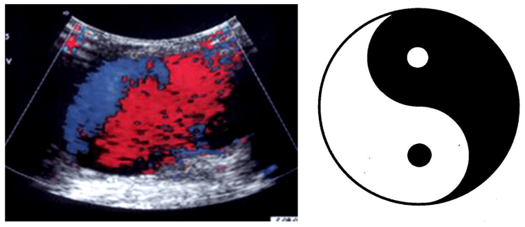 https://abs.twimg.com/emoji/v2/... draggable="false" alt="🔗" title="Link Symbol" aria-label="Emoji: Link Symbol"> https://pocus101.com/dvt"..." title="18 On ultrasound, pseudoaneurysms present as anechoic or hypoechoic images. With Color Doppler, you can see a “Yin-Yang Sign” from the circular motion of blood inside the pseudoaneurysm cavity. Don& #39;t confuse this for a non-compressible vein.https://abs.twimg.com/emoji/v2/... draggable="false" alt="👉" title="Rückhand Zeigefinger nach rechts" aria-label="Emoji: Rückhand Zeigefinger nach rechts">https://abs.twimg.com/emoji/v2/... draggable="false" alt="🔗" title="Link Symbol" aria-label="Emoji: Link Symbol"> https://pocus101.com/dvt"..." class="img-responsive" style="max-width:100%;"/>
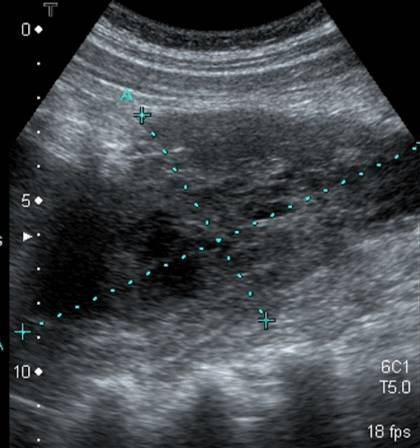 https://abs.twimg.com/emoji/v2/... draggable="false" alt="🔗" title="Link Symbol" aria-label="Emoji: Link Symbol"> https://pocus101.com/dvt"..." title="19 On ultrasound, a groin hematoma will be hypoechoic with some anechoic areas scattered throughout. If you scan the hematoma in the longitudinal view, it becomes obvious that the object is circumscribed and not tubular like a vein or artery.https://abs.twimg.com/emoji/v2/... draggable="false" alt="👉" title="Rückhand Zeigefinger nach rechts" aria-label="Emoji: Rückhand Zeigefinger nach rechts">https://abs.twimg.com/emoji/v2/... draggable="false" alt="🔗" title="Link Symbol" aria-label="Emoji: Link Symbol"> https://pocus101.com/dvt"..." class="img-responsive" style="max-width:100%;"/>
 https://abs.twimg.com/emoji/v2/... draggable="false" alt="🔗" title="Link Symbol" aria-label="Emoji: Link Symbol"> https://pocus101.com/RevBook&q..." title="20 Want to learn even MORE about DVT ultrasound? Check out our newly released #POCUS Board Review Book with an exclusive 20% discount code!https://abs.twimg.com/emoji/v2/... draggable="false" alt="👉" title="Rückhand Zeigefinger nach rechts" aria-label="Emoji: Rückhand Zeigefinger nach rechts">https://abs.twimg.com/emoji/v2/... draggable="false" alt="🔗" title="Link Symbol" aria-label="Emoji: Link Symbol"> https://pocus101.com/RevBook&q..." class="img-responsive" style="max-width:100%;"/>


