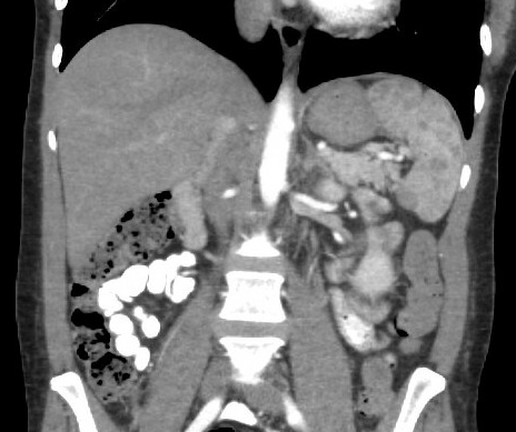
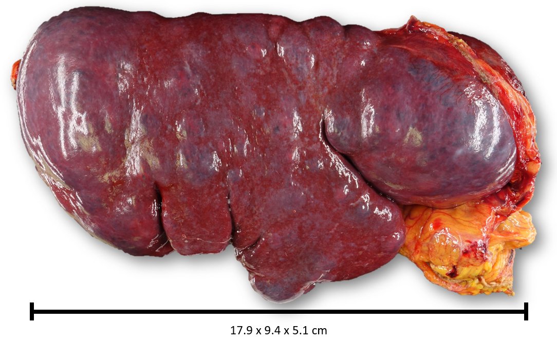
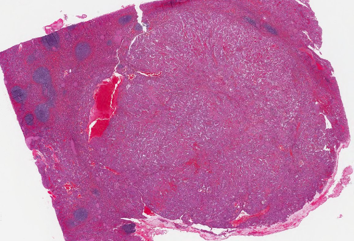
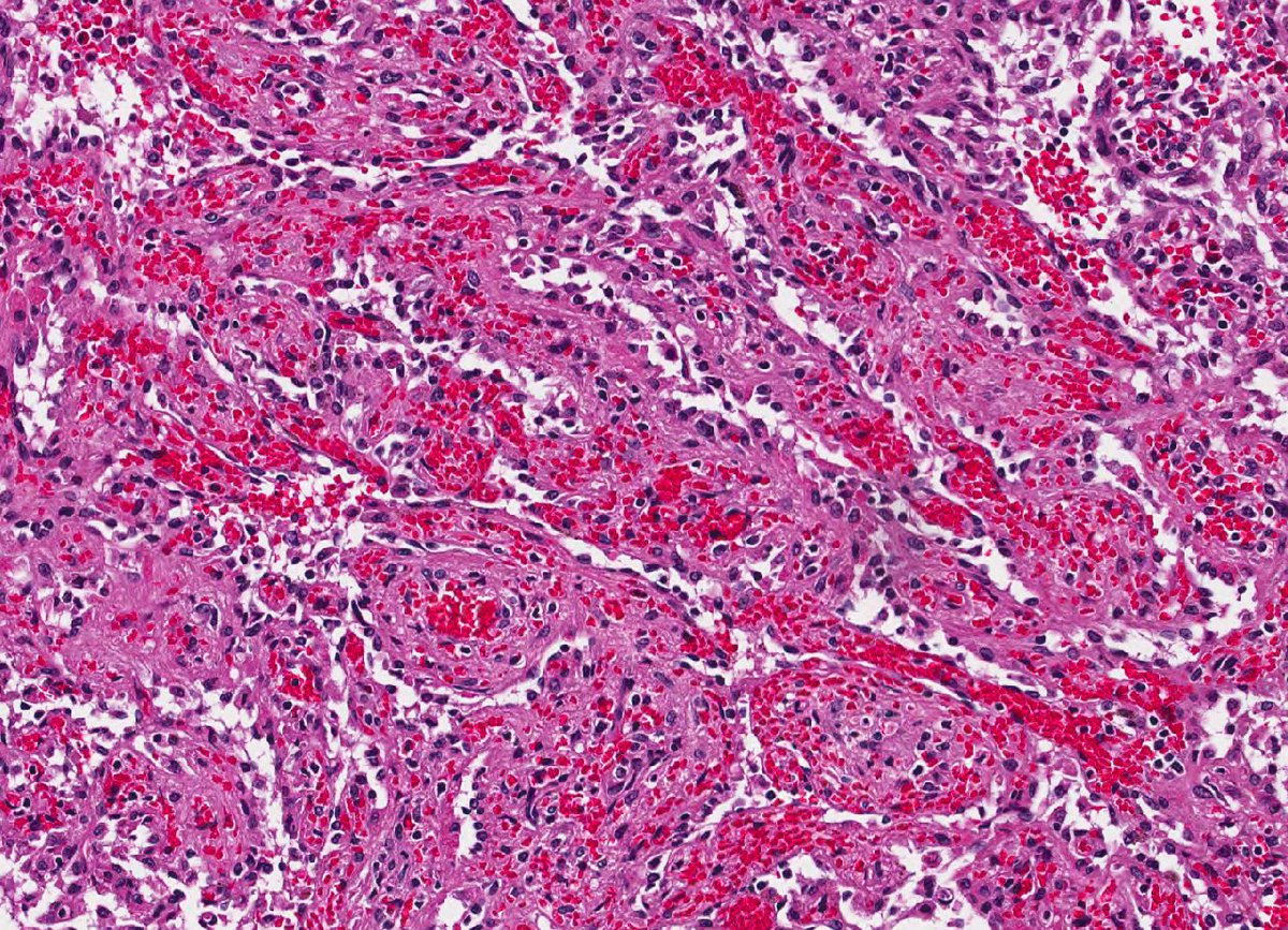
First impression differential?
[SANT: Sclerosing Angiomatous Nodular Tumor]
[SANT: Sclerosing Angiomatous Nodular Tumor]
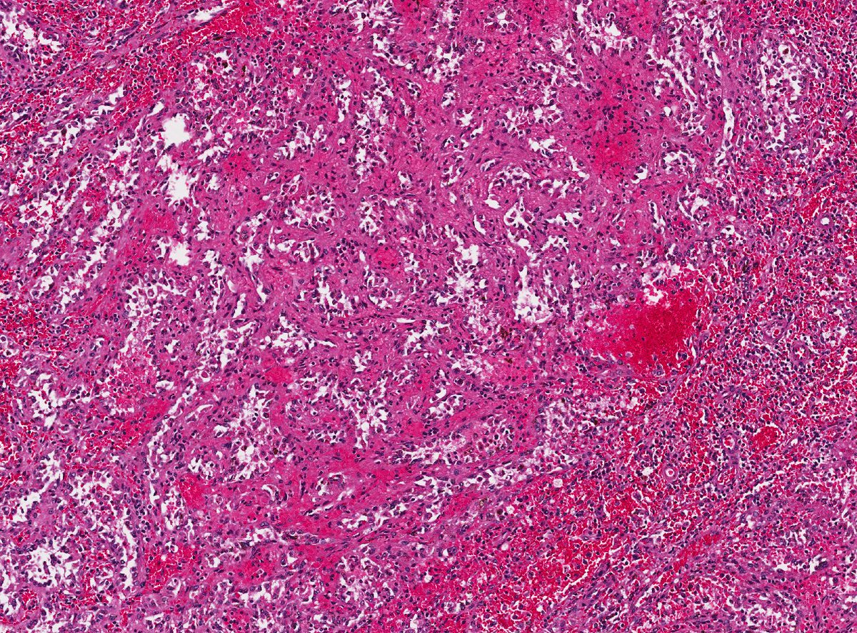
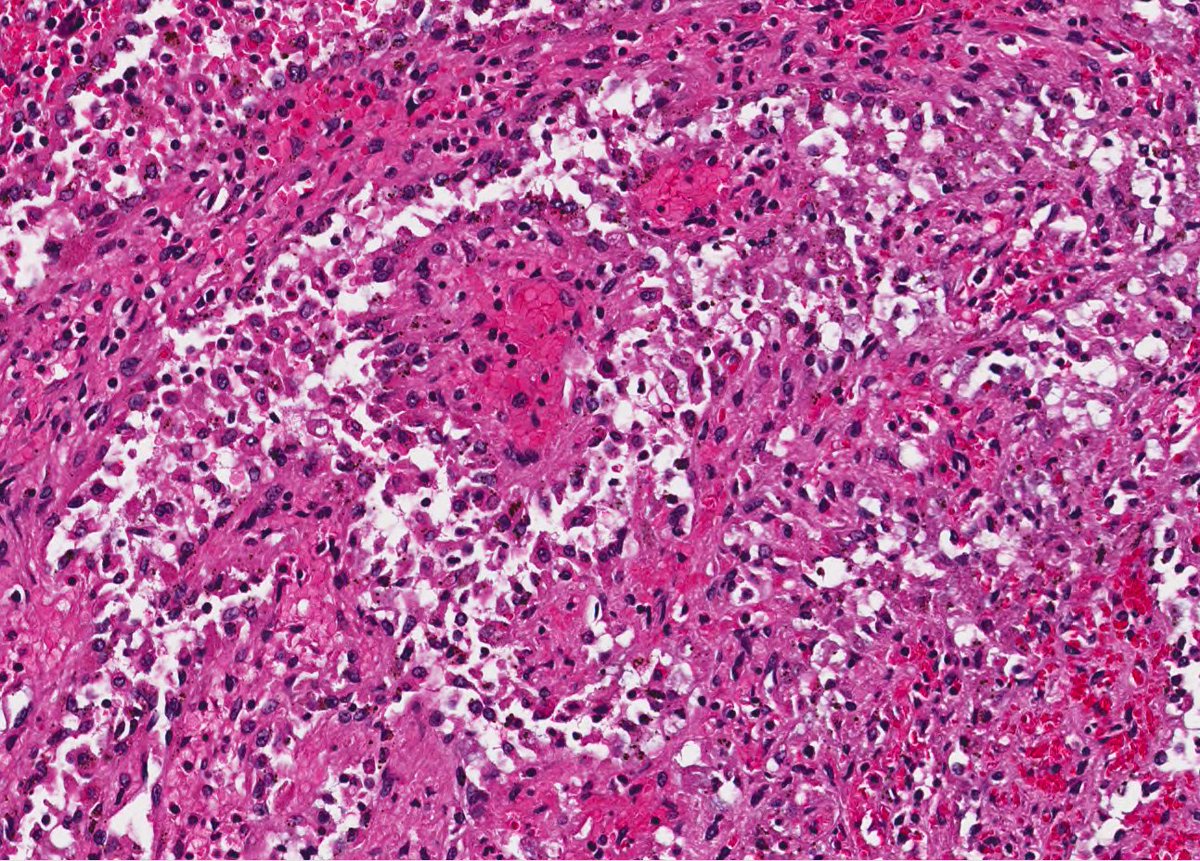
Hard to know without first looking at higher power for atypia (and of course a few stains). First, here& #39;s some higher power.
Nuclear atypia is present -- but minimal. Note the cystic vascular channels filled with appear to be desquamated cells.
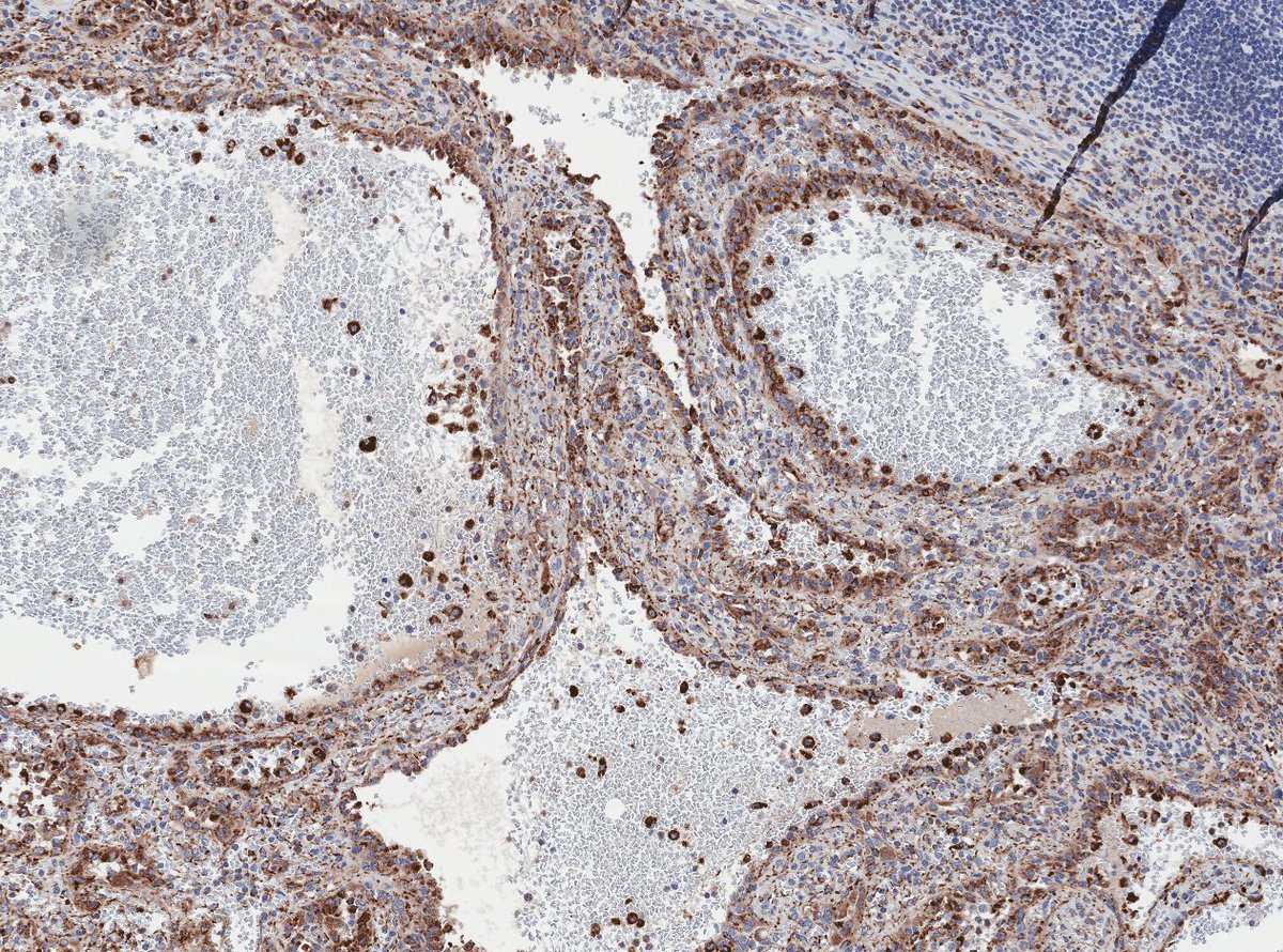
What stain highlights those floating cells (above)?
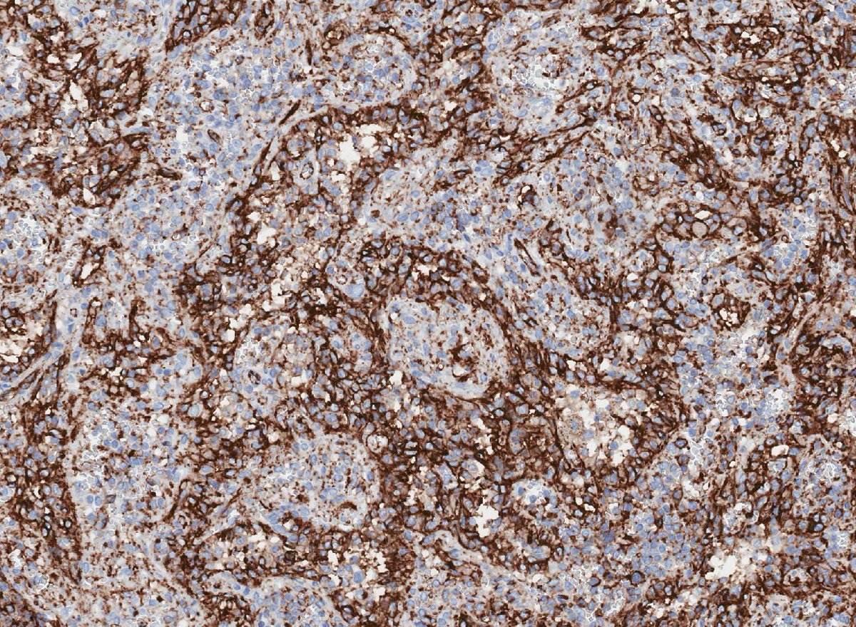
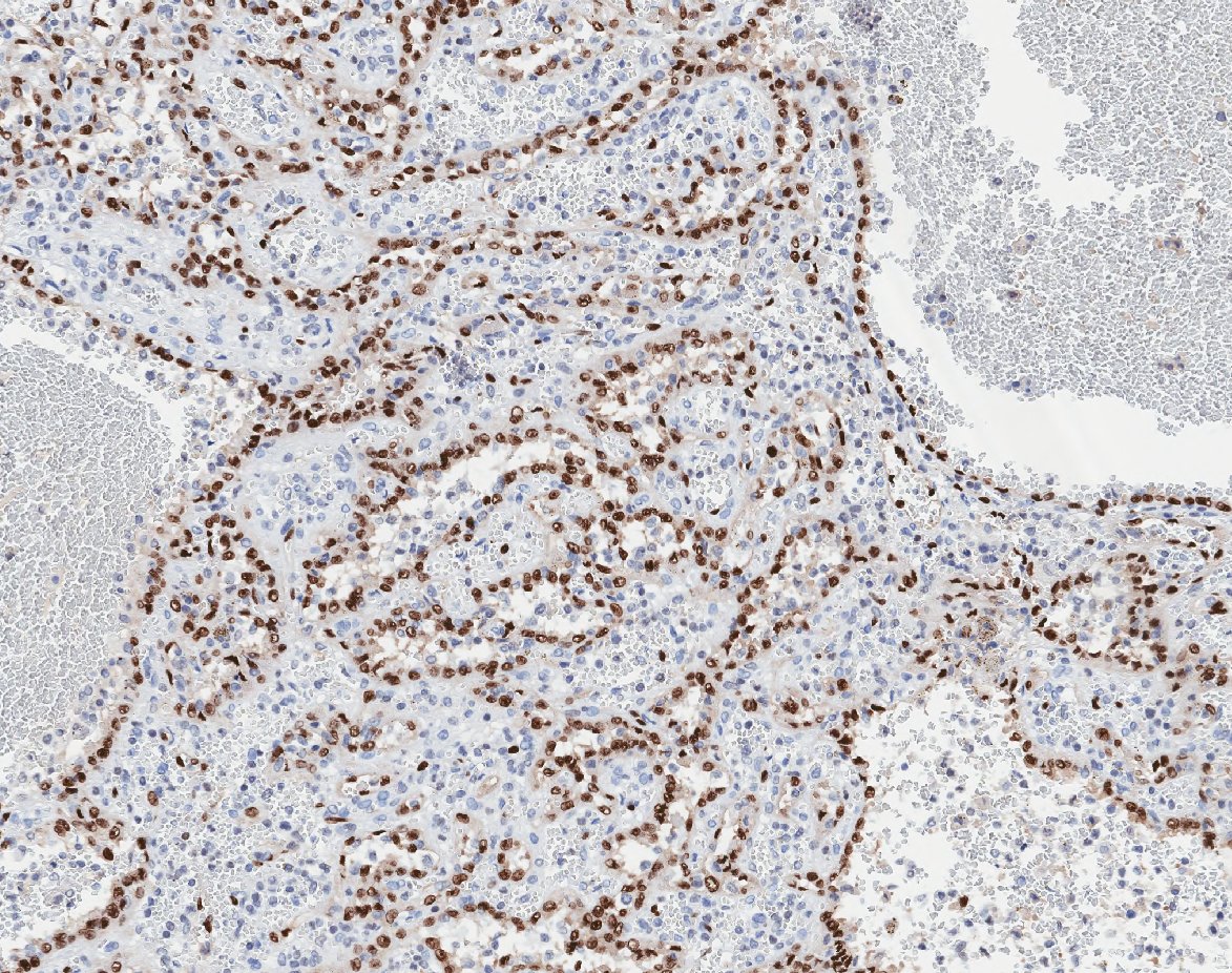
CD68. These are tall lining cells are histiocytic. The flat basal cells are endothelial. Here& #39;s a CD31 (bit messy) and an ERG (infrequently used in this lesion, but it& #39;s so pretty).
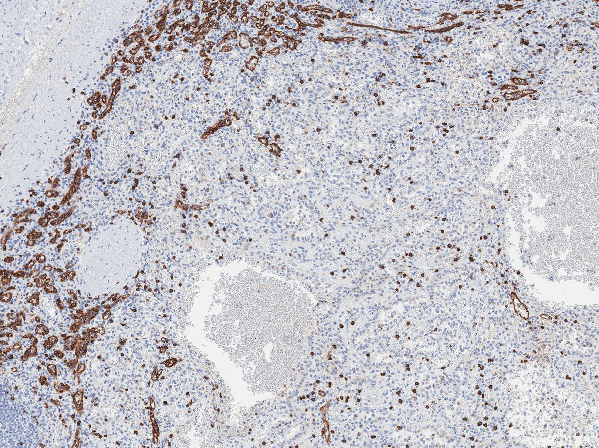
So we have some atypia, but no necrosis, and rare (if any) mitoses. Probably not angiosarc. Here& #39;s a CD8 to show the replacement of normal splenic pulp.
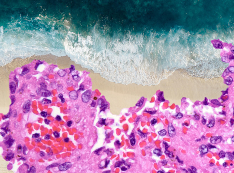
Final diagnosis? Littoral cell angioma. Rare benign splenic vascular neoplasm thought to arise from red pulp sinus lining cells. Littoral means "relating to or situated on the shore of the sea or lake.
As a surfer, I think that& #39;s just awesome.
As a surfer, I think that& #39;s just awesome.

 Read on Twitter