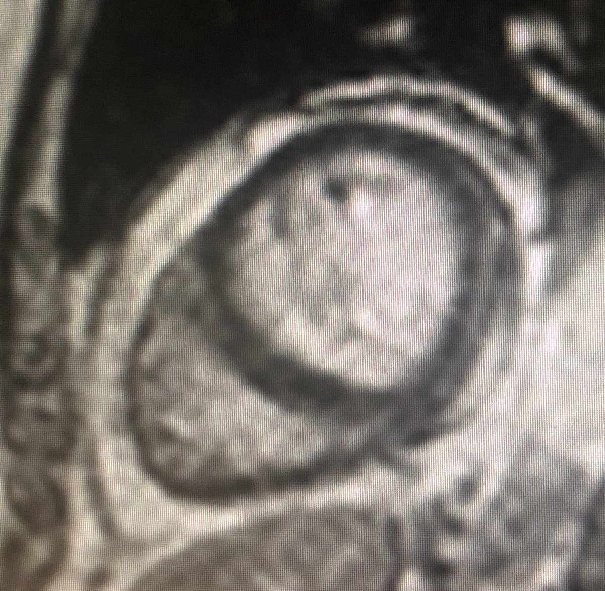
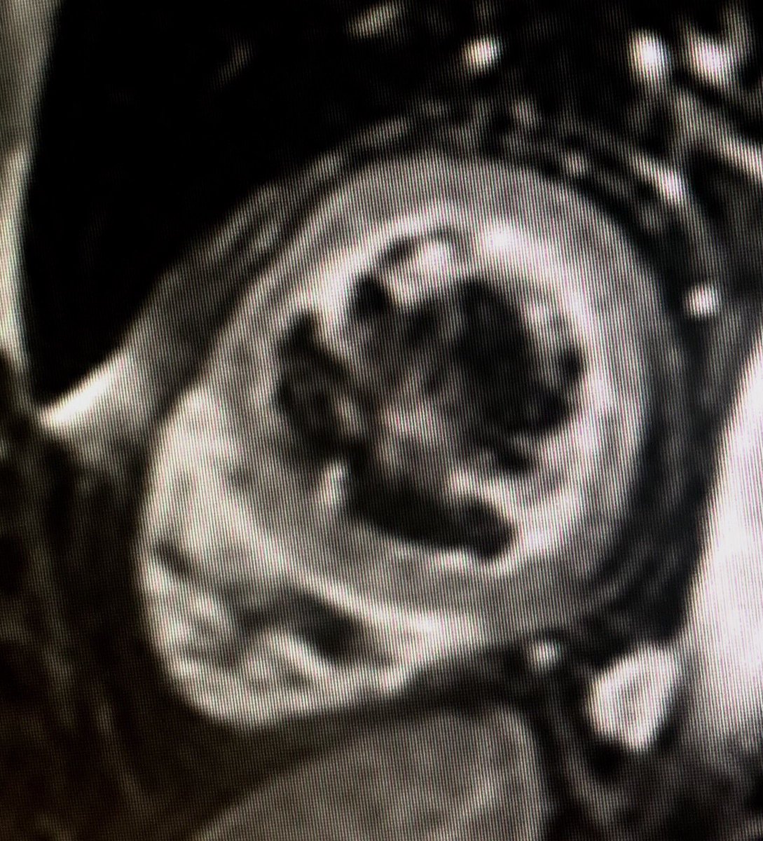
1/9 Tissue characterisation by #WhyCMR in a pt with bright signal in inferior and lateral LV wall on echo suspected of having had an MI but only mild CAD on coronary angio. @scmrorg @chiarabd @purviparwani @PushpaShivaram @AmroAlsaid @danilorenzatti @Kfarooqi @RayRcnita @BSCMR
2/9 Bright signal isodense with fat (epicardial and SC fat) in inferior / inferolateral subepicardial wall (at 3-5 o’clock-disappears with fat suppression sequences - far right) @JStojanovskaMD @DrJenniferCo_Vu @OKhaliqueMD @AScatteia @AKallifatidis @vineetao17 @DmmOsmany
3/9 Ectopic intracardiac fatty foci are not uncommon findings in both healthy & diseased patients.
#YesCCT & #whycmr can detect fat easily within the heart. @vineetao17 @Ahmed43101178 @JoaoLCavalcante @HeartDocSubha @mugander @Doc_Tiger @MasriAhmadMD
#YesCCT & #whycmr can detect fat easily within the heart. @vineetao17 @Ahmed43101178 @JoaoLCavalcante @HeartDocSubha @mugander @Doc_Tiger @MasriAhmadMD
4/9 incidental frequency of cardiac adipose images is about 10% of the patients having #yescct.
However foci of ectopic cardiac adipose tissue are not usually reported even in pathological conditions, mostly if the exam is not specifically focused on the heart.
However foci of ectopic cardiac adipose tissue are not usually reported even in pathological conditions, mostly if the exam is not specifically focused on the heart.
5/9 @RezaEmaminia @ShelbyKuttyMD @EylemLevelt @Sarah_Moharem @DrRyanPDaly @rladeiraslopes @DrFuisz @KimAtianzar @tiffchenMD @cshenoy3 @rooshaparikh @AChoiHeart @onco_cardiology @AkhilNarangMD @heartdockumar @ash71us
6/9 Non-pathological ectopic intramyocardial fat extending from the epicardiac adipose tissue is a relatively common incidental finding during a routine chest or cardiac CT (usu. more frequent in the RV than in the LV).
7/9 Following disease processes may need to be considered when ectopic fat deposits are seen
 https://abs.twimg.com/emoji/v2/... draggable="false" alt="1️⃣" title="Tastenkappe Ziffer 1" aria-label="Emoji: Tastenkappe Ziffer 1">ARVC / ACM
 https://abs.twimg.com/emoji/v2/... draggable="false" alt="2️⃣" title="Tastenkappe Ziffer 2" aria-label="Emoji: Tastenkappe Ziffer 2"> Postmyocardial Infarction Lipomatous Metaplasia (usu. subendocardial)
 https://abs.twimg.com/emoji/v2/... draggable="false" alt="3️⃣" title="Tastenkappe Ziffer 3" aria-label="Emoji: Tastenkappe Ziffer 3"> Postmyocardial inflammation Lipomatous Metaplasia (usu. subepicardial)
 https://abs.twimg.com/emoji/v2/... draggable="false" alt="4️⃣" title="Tastenkappe Ziffer 4" aria-label="Emoji: Tastenkappe Ziffer 4">DCM (usu. mid wall)
8/9  https://abs.twimg.com/emoji/v2/... draggable="false" alt="5️⃣" title="Tastenkappe Ziffer 5" aria-label="Emoji: Tastenkappe Ziffer 5"> Lipomatous Hypertrophy of the Interatrial Septum
 https://abs.twimg.com/emoji/v2/... draggable="false" alt="6️⃣" title="Tastenkappe Ziffer 6" aria-label="Emoji: Tastenkappe Ziffer 6"> Tuberous sclerosis
 https://abs.twimg.com/emoji/v2/... draggable="false" alt="7️⃣" title="Tastenkappe Ziffer 7" aria-label="Emoji: Tastenkappe Ziffer 7"> Lipoma & rarely liposarcoma
 https://abs.twimg.com/emoji/v2/... draggable="false" alt="8️⃣" title="Tastenkappe Ziffer 8" aria-label="Emoji: Tastenkappe Ziffer 8"> others such as Muscular dystrophies, HCM, Fabrys, metabolic disorders

 Read on Twitter


