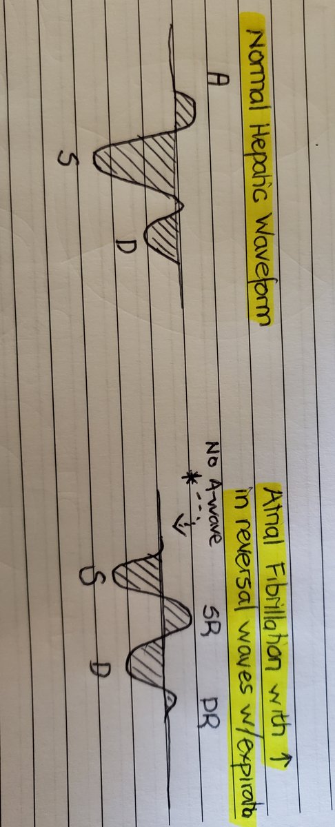
(1/ )There has been an overwhelming interest in the use of hepatic waveforms as a congestive parameter. With social media, somewhat obscure concepts like these are quickly brought to the forefront and implemented in clinical practice. Many are eager to incorporate #VEXUS but...
...a thorough understanding of these waveforms, hemodynamic correlates, and dynamic changes that occur in physiology and disease, is paramount to incorporating this into practice. #shocksquad #tweetorial #VEXUS #showmethewaveform. Here are rules of interpreting hepatic waveforms
(2/13)Rule 1: **Nomenclature, nomenclature, nomenclature! **
That’s right! For us to understand each other we must speak the same language. I have heard these waves being described in a multitude of ways which has brought about considerable confusion and miscommunication.
That’s right! For us to understand each other we must speak the same language. I have heard these waves being described in a multitude of ways which has brought about considerable confusion and miscommunication.
(3/13)Remember understanding nomenclature not only allows you to parlay your information more effectively but it also FORCES you to be more systematic when interpreting these waveforms. We are going to spend some time here so get comfy…
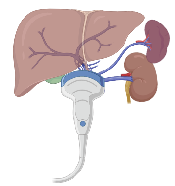
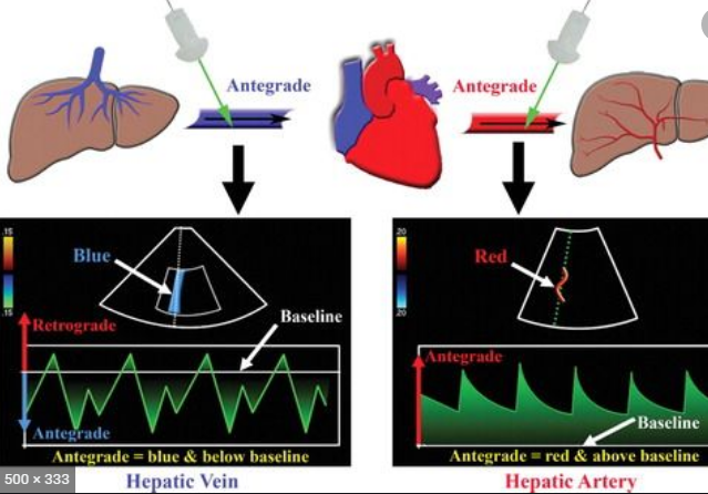
(4/13)*Antegrade vs Retrograde*: this can be in terms of the natural flow within the circulatory system or arbitrarily related to your probe position. RED is towards the probe, BLUE is away.
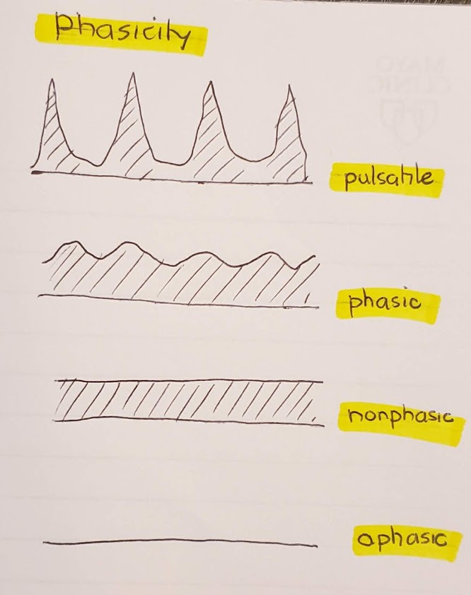
(5/13)*Phasicity* simply indicates degree of fluctuation of the waveform.
Aphasic: no flow at all!
Non-phasic: flow but no fluctuations
Phasic: undulating or fluctuating waveform
Pulsatile: undulating or fluctuating waveform but to a higher higher degree of phasic.
Aphasic: no flow at all!
Non-phasic: flow but no fluctuations
Phasic: undulating or fluctuating waveform
Pulsatile: undulating or fluctuating waveform but to a higher higher degree of phasic.
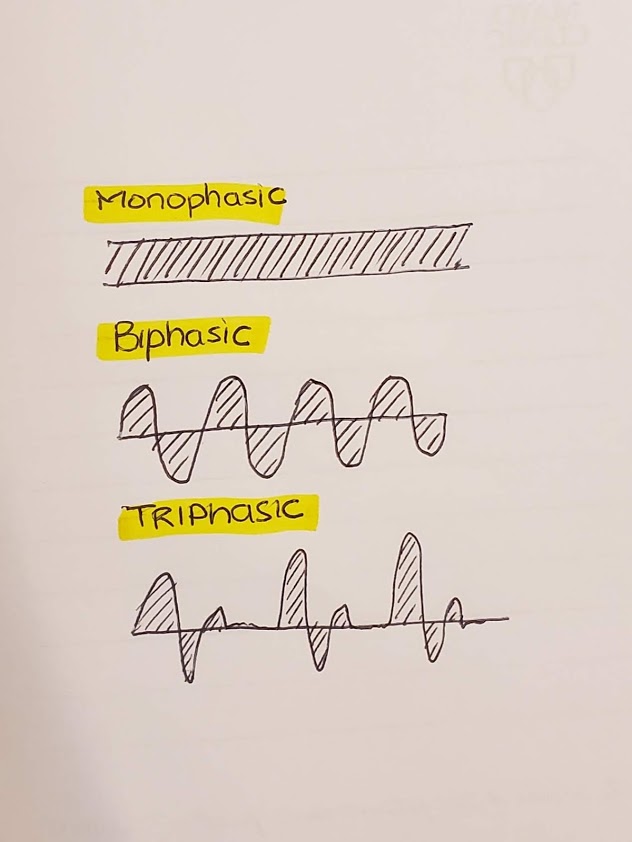
(6/13)*Phase quantification* this is where things get interesting and you will see considerable variability. Simply, a phase occurs when your waveform crosses the baseline. So the phasic and pulsatile flow shown in the above tweet are MONOPHASIC! Bi- and tri-phasic https://abs.twimg.com/emoji/v2/... draggable="false" alt="⬇️" title="Pfeil nach unten" aria-label="Emoji: Pfeil nach unten">
(7/13)*Inflection Quantification* Hmm..the plot thickens. A - biphasic and tri-inflectional, B - tetraphasic and tertra-inflectional. By taking the time to describe these waveforms by inflection points, one will start to understand them better.
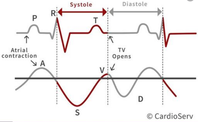
(8/13) Rule 2: **Timing of waveforms w/ cardiac cycle**
“Systole? Diastole? Who cares! I’ve seen so many of these waves I don’t NO EKG leads.” Please don’t be that person. You can be easily misled if you don’t interpret in the context of timing of the cardiac cycle. See below..
“Systole? Diastole? Who cares! I’ve seen so many of these waves I don’t NO EKG leads.” Please don’t be that person. You can be easily misled if you don’t interpret in the context of timing of the cardiac cycle. See below..
(9/13) Rule 3: **Timing of mechanical events**
Atrial contraction: a-wave
Atrial relaxation: downslope of a-wave
TV valve closure:
RV Contraction: S- wave
Atrial Overfilling: upsloping of S-wave (may go over baseline)
RV relaxation: D-wave
Atrial contraction: a-wave
Atrial relaxation: downslope of a-wave
TV valve closure:
RV Contraction: S- wave
Atrial Overfilling: upsloping of S-wave (may go over baseline)
RV relaxation: D-wave
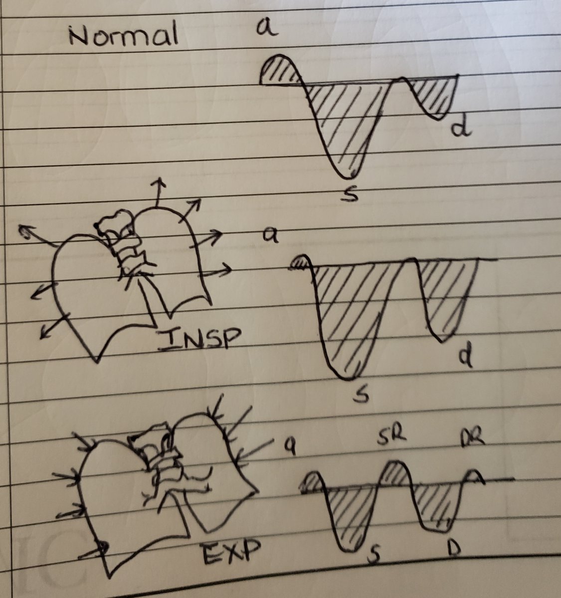
(10/13) Rule 4: ** Changes with Breathing **
With inspiration antegrade flow (S and D waves increase), With expiration the reversal waves increase (SR and DR).
With inspiration antegrade flow (S and D waves increase), With expiration the reversal waves increase (SR and DR).
(11/13) Remember these rules well! In the next installment we will go over
- Changes with PPV
- Liver parenchymal disease
- Changes in A wave in atrial arrhythmia
- S and D-wave morphology, changes and reversal
- Constriction, restriction
-Special circumstances
- Changes with PPV
- Liver parenchymal disease
- Changes in A wave in atrial arrhythmia
- S and D-wave morphology, changes and reversal
- Constriction, restriction
-Special circumstances
(12/13) Interpreting these waveforms can add a tremendous amount of clinical information when combined with the clinical picture. We have a tendency to flock to new technology and forget the basics. I& #39;ve seen an over reliance on POCUS...
(13/13) Stay tuned for the next installment. Where we will see how compliance, TR, RV dysfunction, constriction, and restriction come into play@PittIMPOCUS @sono @POCUS_Society @siddharth_dugar @msiuba @load_dependent @ThinkingCC @iceman_ex @khaycock2 @Yale_EUS @IM_POCUS @NephroP

 Read on Twitter
 " title="(6/13)*Phase quantification* this is where things get interesting and you will see considerable variability. Simply, a phase occurs when your waveform crosses the baseline. So the phasic and pulsatile flow shown in the above tweet are MONOPHASIC! Bi- and tri-phasichttps://abs.twimg.com/emoji/v2/... draggable="false" alt="⬇️" title="Pfeil nach unten" aria-label="Emoji: Pfeil nach unten">" class="img-responsive" style="max-width:100%;"/>

