Hey #MedStudentTwitter #neurology residents, another #EndNeurophobia #MedEd #tweetorial to celebrate joining the @CPSolvers team!
CORTICAL REGIONS AND STROKE SYNDROMES
cc: @Tracey1milligan @MadSattinJ @CrystalYeoMDPhD
@DxRxEdu @MedTweetorials
CORTICAL REGIONS AND STROKE SYNDROMES
cc: @Tracey1milligan @MadSattinJ @CrystalYeoMDPhD
@DxRxEdu @MedTweetorials
But first a moment of silence for
#RayshardBrooks
#BreonnaTaylor
#GeorgeFloyd
#AhmaudArbery
.
.
.
.
.
.
.
.
.
.
#RayshardBrooks
#BreonnaTaylor
#GeorgeFloyd
#AhmaudArbery
.
.
.
.
.
.
.
.
.
.
Grateful for https://abs.twimg.com/emoji/v2/... draggable="false" alt="❤️" title="Rotes Herz" aria-label="Emoji: Rotes Herz"> for #EndNeurophobia but hope you read rest of my feed, which seeks to amplify voices I’m learning from on the path to being an antiracist ally.
to learn more, follow/learn from #BlackMedTwitter #BlackintheIvory
listen @thepraxispod @thenocturnists
read @dribram
to learn more, follow/learn from #BlackMedTwitter #BlackintheIvory
listen @thepraxispod @thenocturnists
read @dribram
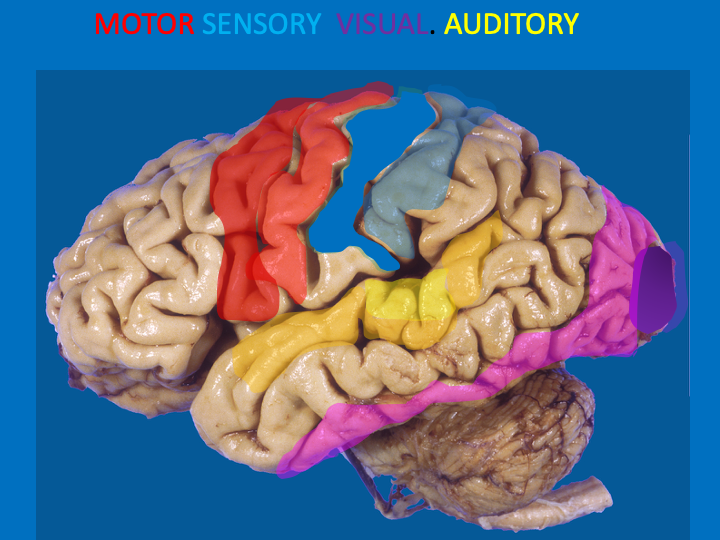
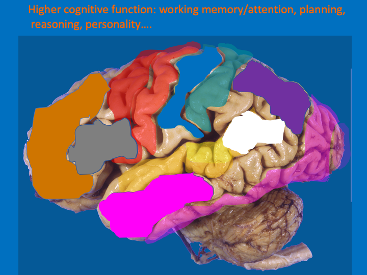
If you can identify 4 regions of cortex–1 motor/3 sensory–you can deduce the rest by what they lay between
4 regions=
Motor cortex (precentral gyrus)
Somatosensory cortex (postcentral gyrus)
Visual cortex (posterior occipital lobe)
Auditory cortex (superior temporal gyrus)
4 regions=
Motor cortex (precentral gyrus)
Somatosensory cortex (postcentral gyrus)
Visual cortex (posterior occipital lobe)
Auditory cortex (superior temporal gyrus)
These regions are primary regions and are surrounded by unimodal association cortex, meaning higher level processing of the same modality of information
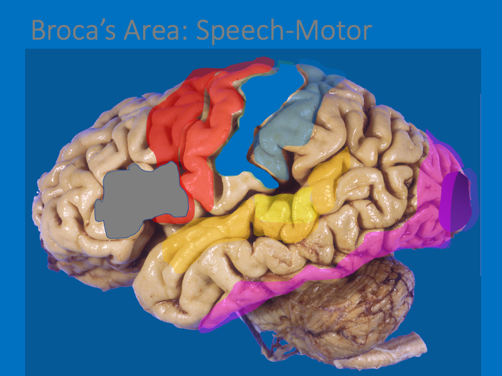
What would logically be @ junction of auditory &motor?
BROCA’S AREA: for speech production; speech=using motor actions to create sound.
Broca’s area is in the inferior frontal gyrus, on the left in all right handers and most left handers.
BROCA’S AREA: for speech production; speech=using motor actions to create sound.
Broca’s area is in the inferior frontal gyrus, on the left in all right handers and most left handers.
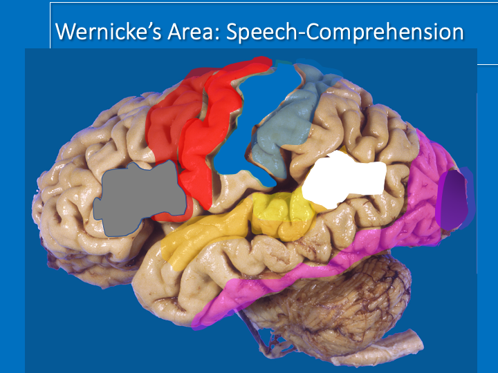
What would logically be @ junction of auditory & parietal?
(as we’ll see parietal plays a role in attention to the outside world)
WERNICKE’s AREA for speech comprehension: attention to sounds in the outside world!
(as we’ll see parietal plays a role in attention to the outside world)
WERNICKE’s AREA for speech comprehension: attention to sounds in the outside world!
A lesion of Broca’s area causes difficulty speaking: anything from word finding difficulties to mutism depending on lesion
A lesion of Wernicke’s area affects speech comprehension (and the patient’s speech is altered as well since they can’t understand what they’re saying)
A lesion of Wernicke’s area affects speech comprehension (and the patient’s speech is altered as well since they can’t understand what they’re saying)
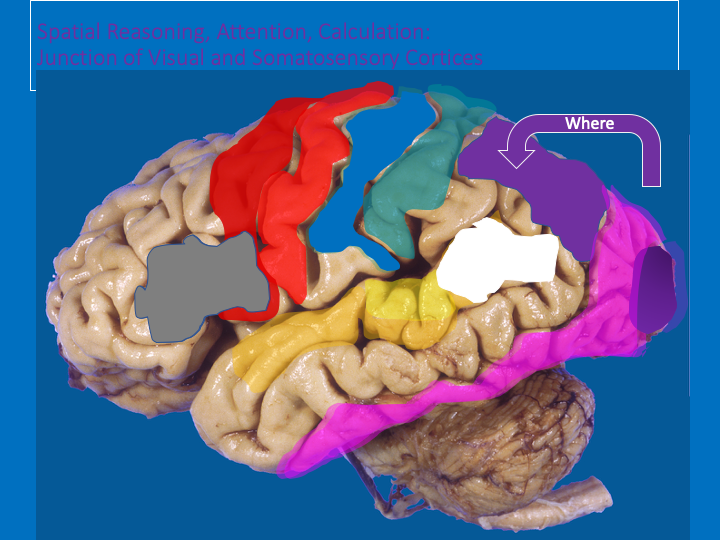
At the junction of visual and somatosensory= WHERE pathway (dorsal visual stream)
The regions that determine WHERE things are and WHERE we are
Parietal lobe involved in attention, spatial reasoning, mathematics…
Lesions of RIGHT parietal lobe can cause hemi-spatial neglect
The regions that determine WHERE things are and WHERE we are
Parietal lobe involved in attention, spatial reasoning, mathematics…
Lesions of RIGHT parietal lobe can cause hemi-spatial neglect
The ventral visual stream is the WHAT pathway, which goes into the temporal lobe. The temporal lobe is ideally situated to receive auditory, visual, somatosensory and smell information to determine WHAT things are=RECOGNITION MEMORY (hippocampus and surrounding regions)
The rest of the frontal lobe is involved in higher order cognitive functions: working memory, planning, reasoning, personality…
So now that we’ve mapped the cortex, we can understand different stroke syndromes that affect it!
So now that we’ve mapped the cortex, we can understand different stroke syndromes that affect it!
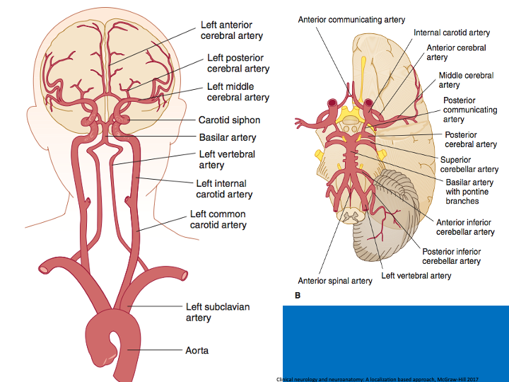
The brain receives its blood supply from the
ANTERIOR circulation (Carotid->MCA and ACA) and
POSTERIOR circulation (Vertebro-basilar system to cerebellar arteries and PCA).
For more on the cerebellar arteries that supply brainstem/cerebellum see prior tweetorials
ANTERIOR circulation (Carotid->MCA and ACA) and
POSTERIOR circulation (Vertebro-basilar system to cerebellar arteries and PCA).
For more on the cerebellar arteries that supply brainstem/cerebellum see prior tweetorials
The posterior and anterior circulation are linked by the POSTERIOR COMMUNICATING arteries (Pcomms) and the ACAs by the ANTERIOR COMMUNICATING arteries (AComms). This makes a complete circle, the CIRCLE OF WILLIS
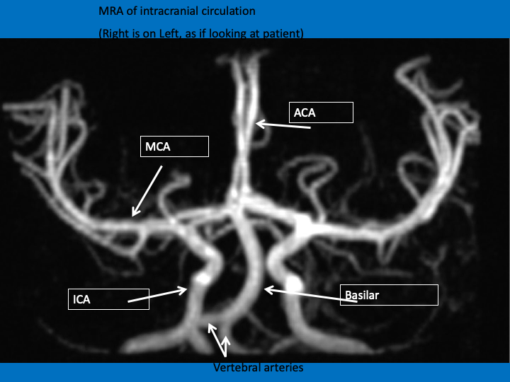
Here’s how we often see the cerebrovascular system: MRA
Here’s how we often see the cerebrovascular system: MRA
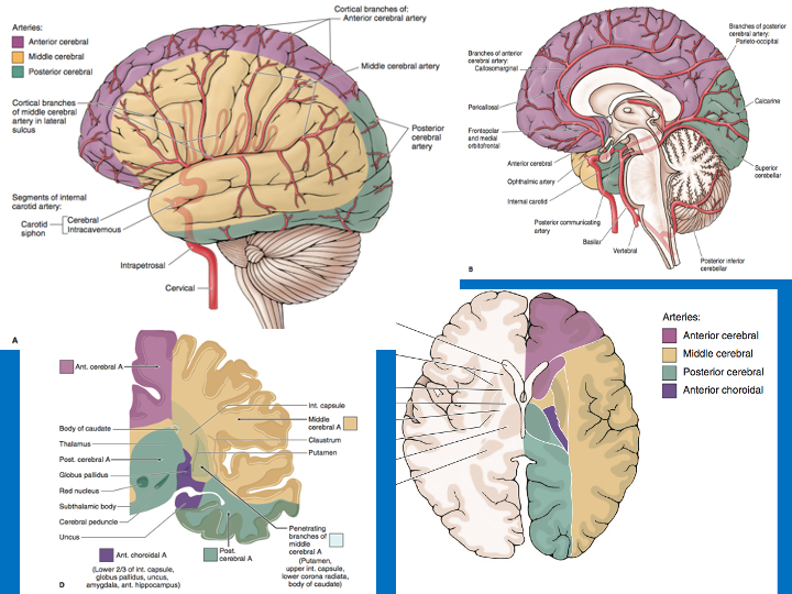
And here are the cerebral vascular territories
MCA: lateral surface of frontal, temporal, parietal lobes as well as much of subcortical white/gray matter
ACA: Superior and medial frontal and parietal lobes; anterior frontal lobe
PCA: occipital, inferior temporal, and thalamus
MCA: lateral surface of frontal, temporal, parietal lobes as well as much of subcortical white/gray matter
ACA: Superior and medial frontal and parietal lobes; anterior frontal lobe
PCA: occipital, inferior temporal, and thalamus
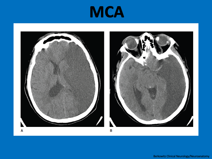
CT w/full territory MCA stroke. This will cause
Contralateral hemiplegia(motor ctx)/hemisensory loss (somatosensory ctx)/homonymous hemianopia (optic radiations)
Language deficit (if left)/Neglect (if right)
Strokes affecting just branches of MCA can cause lesser/fewer sx
Contralateral hemiplegia(motor ctx)/hemisensory loss (somatosensory ctx)/homonymous hemianopia (optic radiations)
Language deficit (if left)/Neglect (if right)
Strokes affecting just branches of MCA can cause lesser/fewer sx
On prior, Note regions NOT affected on CT as they are supplied by
ACA (anteriorly; &caudate supplied by recurrent artery of Huebner, an ACA branch)
and PCA (posteriorly)
ACA (anteriorly; &caudate supplied by recurrent artery of Huebner, an ACA branch)
and PCA (posteriorly)
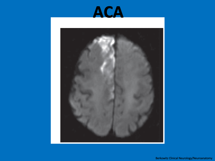
MRI w/ACA stroke
Recall that homunculus=leg medial, arm superior/lateral, face most lateral
So ACA stroke can cause contralateral leg>arm weakness &cognitive changes
I’ve only seen a few ACA strokes but countless MCA, PCA strokes–perhaps the fluid dynamics/geometry—thoughts?
Recall that homunculus=leg medial, arm superior/lateral, face most lateral
So ACA stroke can cause contralateral leg>arm weakness &cognitive changes
I’ve only seen a few ACA strokes but countless MCA, PCA strokes–perhaps the fluid dynamics/geometry—thoughts?
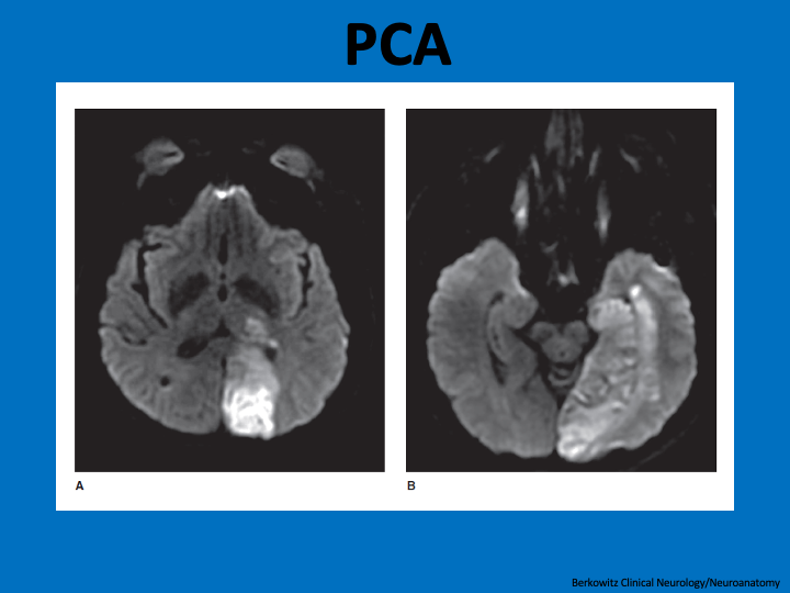
This MRI shows a PCA stroke:
PCA strokes cause contralateral visual field deficits and can cause amnesia if hippocampus affected. Note how PCA includes inferior/medial temporal lobe and thalamus, not just visual cortex
PCA strokes cause contralateral visual field deficits and can cause amnesia if hippocampus affected. Note how PCA includes inferior/medial temporal lobe and thalamus, not just visual cortex
To close, Some gourmet vascular anatomy:
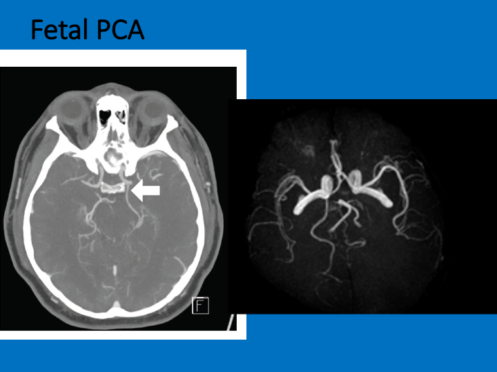
FETAL PCA=normal variant of PCA coming off ANTERIOR circulation
No clinical relevance UNLESS PCA stroke on side of fetal PCA 2/2 severe carotid stenosis=would mean carotid symptomatic (normally could only be case w/ACA or MCA stroke)
FETAL PCA=normal variant of PCA coming off ANTERIOR circulation
No clinical relevance UNLESS PCA stroke on side of fetal PCA 2/2 severe carotid stenosis=would mean carotid symptomatic (normally could only be case w/ACA or MCA stroke)
Artery of Percheron stroke
Rarely both thalami supplied by single artery.
This is one of the rare strokes that can present with ALTERED MENTAL STATUS without obvious focal features. Others include: inf division rMCA, ACA (if motor cortex spared), PCA, diffuse emboli
Rarely both thalami supplied by single artery.
This is one of the rare strokes that can present with ALTERED MENTAL STATUS without obvious focal features. Others include: inf division rMCA, ACA (if motor cortex spared), PCA, diffuse emboli
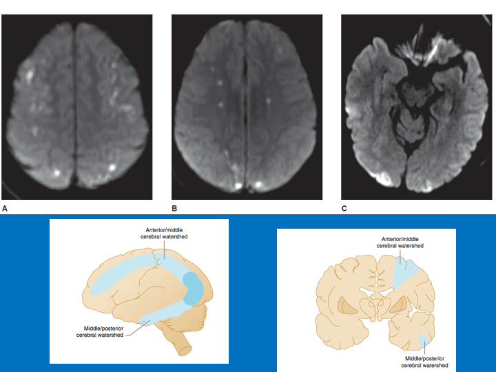
Watershed/borderzone=regions@ borders of territories
ACA/MCA MCA/PCA=most common
We& #39;re taught borderzone strokes=hypotension mechanism
But can be EMBOLIC: smallest possible emboli end up in the end-arterial territories
For discussion see https://pubmed.ncbi.nlm.nih.gov/9823834/ ">https://pubmed.ncbi.nlm.nih.gov/9823834/&...
ACA/MCA MCA/PCA=most common
We& #39;re taught borderzone strokes=hypotension mechanism
But can be EMBOLIC: smallest possible emboli end up in the end-arterial territories
For discussion see https://pubmed.ncbi.nlm.nih.gov/9823834/ ">https://pubmed.ncbi.nlm.nih.gov/9823834/&...
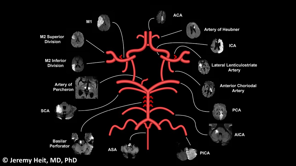
In sum, @JeremyHeitMDPHD & #39;s AMAZING figure
What should I cover next #MedTwitter?
#Endneurophobia https://abs.twimg.com/emoji/v2/... draggable="false" alt="🧠" title="Gehirn" aria-label="Emoji: Gehirn">
 https://abs.twimg.com/emoji/v2/... draggable="false" alt="❤️" title="Rotes Herz" aria-label="Emoji: Rotes Herz">
What should I cover next #MedTwitter?
#Endneurophobia

 Read on Twitter


 https://abs.twimg.com/emoji/v2/... draggable="false" alt="❤️" title="Rotes Herz" aria-label="Emoji: Rotes Herz">" title="In sum, @JeremyHeitMDPHD & #39;s AMAZING figureWhat should I cover next #MedTwitter? #Endneurophobia https://abs.twimg.com/emoji/v2/... draggable="false" alt="🧠" title="Gehirn" aria-label="Emoji: Gehirn">https://abs.twimg.com/emoji/v2/... draggable="false" alt="❤️" title="Rotes Herz" aria-label="Emoji: Rotes Herz">" class="img-responsive" style="max-width:100%;"/>


