What do you know about the catheter walk? Can you spot the difference?
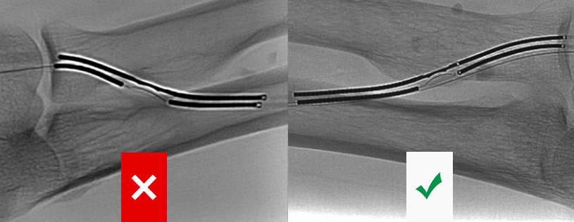
The difference in electrode deflection from #1 to #2 can be minimal but one will result in successful creation, the other in failed creation. I& #39;ll use my crude, homemade model to try to explain:
In example #1, the arterial and venous electrodes are well aligned
With proper alignment, the arterial ceramic footplate will significantly deflect the venous electrode when walked
In example #2, the electrodes are askew
Although you may see some deflection with catheter walking, it will not be nearly as obvious and/or convincing. If you fire in an askew position, the venous electrode will miss the ceramic footplate and you won& #39;t get AVF creation
What happens if you don& #39;t recognize the catheter misalignment?!
As we saw on the model, the electrodes won& #39;t connect and fistula creation will be unsuccessful. Can you spot the difference? (Don& #39;t worry if your answer is  https://abs.twimg.com/emoji/v2/... draggable="false" alt="🤷" title="Person shrugging" aria-label="Emoji: Person shrugging">
 https://abs.twimg.com/emoji/v2/... draggable="false" alt="🤷♀️" title="Achselzuckende Frau" aria-label="Emoji: Achselzuckende Frau">)
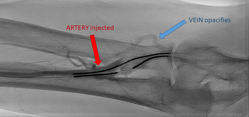
#1 - Electrodes misaligned and do NOT coapt
#1 - Electrodes misaligned and do NOT coapt
#2 - Proper alignment, fistula creation successful  https://abs.twimg.com/emoji/v2/... draggable="false" alt="🙌" title="Raising hands" aria-label="Emoji: Raising hands">
Again, don& #39;t worry if you couldn& #39;t be sure of the difference. If you& #39;re aligned, you know and everyone& #39;s all like
 https://abs.twimg.com/emoji/v2/... draggable="false" alt="🙌" title="Raising hands" aria-label="Emoji: Raising hands">
 https://abs.twimg.com/emoji/v2/... draggable="false" alt="👏" title="Applaus-Zeichen" aria-label="Emoji: Applaus-Zeichen">
 https://abs.twimg.com/emoji/v2/... draggable="false" alt="👍" title="Thumbs up" aria-label="Emoji: Thumbs up">
  https://abs.twimg.com/emoji/v2/... draggable="false" alt="🤜" title="Nach rechts zeigende Faust" aria-label="Emoji: Nach rechts zeigende Faust">
 https://abs.twimg.com/emoji/v2/... draggable="false" alt="🤛" title="Nach links zeigende Faust" aria-label="Emoji: Nach links zeigende Faust">
And if you& #39;re not aligned, the mood is more
 https://abs.twimg.com/emoji/v2/... draggable="false" alt="🤔" title="Denkendes Gesicht" aria-label="Emoji: Denkendes Gesicht">
Buuut, you& #39;re going to confirm creation with contrast injection anyway so that will tell you for sure
And if you& #39;re not aligned, the mood is more
Buuut, you& #39;re going to confirm creation with contrast injection anyway so that will tell you for sure
Last additions to this thread (can you figure out when I have academic time  https://abs.twimg.com/emoji/v2/... draggable="false" alt="😂" title="Gesicht mit Freudentränen" aria-label="Emoji: Gesicht mit Freudentränen">)
How does misalignment happen?
How does misalignment happen?
The first possibility is you inserted the electrodes wrong.
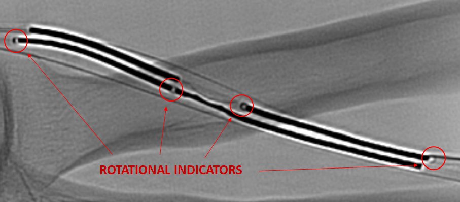
Me: "This is what your rotational indicators are for!"
You: "What the heck are rotational indicators?!"
Me: "This is what your rotational indicators are for!"
You: "What the heck are rotational indicators?!"
They are little cutout squares on the electrodes that look thin edged when well positioned and a solid dark square when in the wrong position. The newer generation device has loads of rotational indicators to help you out  https://abs.twimg.com/emoji/v2/... draggable="false" alt="🙏" title="Folded hands" aria-label="Emoji: Folded hands">
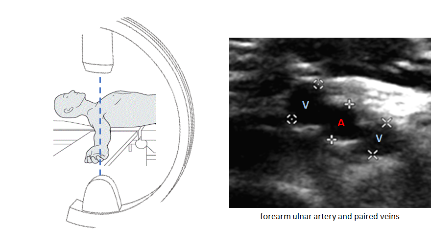
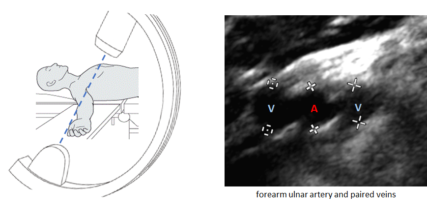
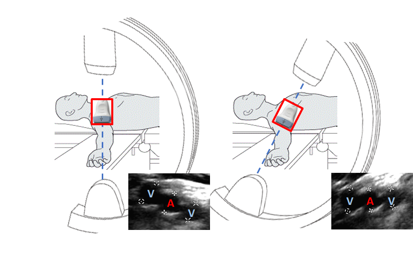
The second option, and more subtle technical issue is that you haven& #39;t obtained the true "widest view." If your image intensifier is not angled properly to place the vessels in the "widest view," your catheters will not align properly.
After trial and error (and lots of squinting), I& #39;ve found the easiest way to get the proper image intensifier angle is to use US first to get the vessels in plane and THEN line the image intensifier up with the US angle.

 Read on Twitter
 https://abs.twimg.com/emoji/v2/... draggable="false" alt="👏" title="Applaus-Zeichen" aria-label="Emoji: Applaus-Zeichen">https://abs.twimg.com/emoji/v2/... draggable="false" alt="👍" title="Thumbs up" aria-label="Emoji: Thumbs up"> https://abs.twimg.com/emoji/v2/... draggable="false" alt="🤜" title="Nach rechts zeigende Faust" aria-label="Emoji: Nach rechts zeigende Faust">https://abs.twimg.com/emoji/v2/... draggable="false" alt="🤛" title="Nach links zeigende Faust" aria-label="Emoji: Nach links zeigende Faust">And if you& #39;re not aligned, the mood is more https://abs.twimg.com/emoji/v2/... draggable="false" alt="🤔" title="Denkendes Gesicht" aria-label="Emoji: Denkendes Gesicht">Buuut, you& #39;re going to confirm creation with contrast injection anyway so that will tell you for sure" title="Again, don& #39;t worry if you couldn& #39;t be sure of the difference. If you& #39;re aligned, you know and everyone& #39;s all likehttps://abs.twimg.com/emoji/v2/... draggable="false" alt="🙌" title="Raising hands" aria-label="Emoji: Raising hands">https://abs.twimg.com/emoji/v2/... draggable="false" alt="👏" title="Applaus-Zeichen" aria-label="Emoji: Applaus-Zeichen">https://abs.twimg.com/emoji/v2/... draggable="false" alt="👍" title="Thumbs up" aria-label="Emoji: Thumbs up"> https://abs.twimg.com/emoji/v2/... draggable="false" alt="🤜" title="Nach rechts zeigende Faust" aria-label="Emoji: Nach rechts zeigende Faust">https://abs.twimg.com/emoji/v2/... draggable="false" alt="🤛" title="Nach links zeigende Faust" aria-label="Emoji: Nach links zeigende Faust">And if you& #39;re not aligned, the mood is more https://abs.twimg.com/emoji/v2/... draggable="false" alt="🤔" title="Denkendes Gesicht" aria-label="Emoji: Denkendes Gesicht">Buuut, you& #39;re going to confirm creation with contrast injection anyway so that will tell you for sure" class="img-responsive" style="max-width:100%;"/>
 " title="They are little cutout squares on the electrodes that look thin edged when well positioned and a solid dark square when in the wrong position. The newer generation device has loads of rotational indicators to help you out https://abs.twimg.com/emoji/v2/... draggable="false" alt="🙏" title="Folded hands" aria-label="Emoji: Folded hands">" class="img-responsive" style="max-width:100%;"/>
