What do we have here? A nice case, poll (below) in thread, and explanation to follow for all the #EchoFirst enthusiasts. #FellowsFirst #ACCFIT #cardiotwitter #ASE360
What do you think is the mechanism for these 2D and Doppler findings? @DavidWienerMD @iamritu @purviparwani @ErinMichos @DrMarthaGulati @HeartDocSharon @HeartOTXHeartMD @mmamas1973 @onco_cardiology @kgzimmerman @rajdoc2005 @DocStrom
1/  https://abs.twimg.com/emoji/v2/... draggable="false" alt="💡" title="Elektrische Glühbirne" aria-label="Emoji: Elektrische Glühbirne">There is constriction, but specifically effusive-constrictive pericarditis.
 https://abs.twimg.com/emoji/v2/... draggable="false" alt="👉" title="Rückhand Zeigefinger nach rechts" aria-label="Emoji: Rückhand Zeigefinger nach rechts"> #tweetorial here:
 https://abs.twimg.com/emoji/v2/... draggable="false" alt="🔶" title="Große orangene Raute" aria-label="Emoji: Große orangene Raute">To understand #EchoFirst findings in pericardial disease, must go back to basics
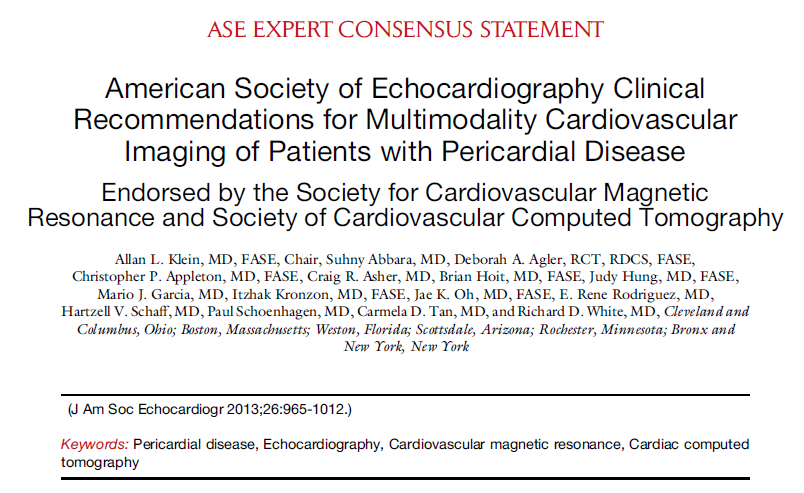
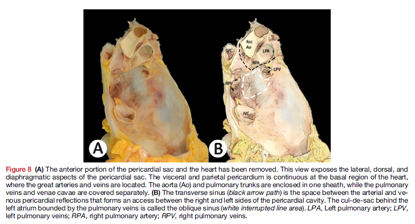
 https://abs.twimg.com/emoji/v2/... draggable="false" alt="🔶" title="Große orangene Raute" aria-label="Emoji: Große orangene Raute">Remember that superior portion of LA and pulmonary veins are extra-pericardial structures.
2/ In constrictive pericarditis (CP) or cardiac tamponade (CT), inspiratory https://abs.twimg.com/emoji/v2/... draggable="false" alt="⬇️" title="Pfeil nach unten" aria-label="Emoji: Pfeil nach unten">of intra-thoracic pressure & in PVeins is not transmitted to LA or LV. This dissociation leads to
 https://abs.twimg.com/emoji/v2/... draggable="false" alt="⬇️" title="Pfeil nach unten" aria-label="Emoji: Pfeil nach unten">PVein-LA gradient and
  https://abs.twimg.com/emoji/v2/... draggable="false" alt="⬇️" title="Pfeil nach unten" aria-label="Emoji: Pfeil nach unten">LV filling, demonstrated by an inspiratory
  https://abs.twimg.com/emoji/v2/... draggable="false" alt="⬇️" title="Pfeil nach unten" aria-label="Emoji: Pfeil nach unten">in mitral inflow velocity by >25%.
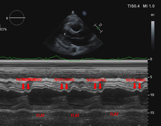
3/ In CP. early diastolic septal "bounce" is noted in 2D and M-mode. M-mode also reveals rapid relaxation of posterior wall followed by "flattening" of motion throughout diastole. Ventricular interdependence seen in CP and CT, septum shifts L in inspiration and R in expiration.
4/ In restriction, tissue Doppler is abnormal ( https://abs.twimg.com/emoji/v2/... draggable="false" alt="⬇️" title="Pfeil nach unten" aria-label="Emoji: Pfeil nach unten">e& #39; and
  https://abs.twimg.com/emoji/v2/... draggable="false" alt="⬆️" title="Pfeil nach oben" aria-label="Emoji: Pfeil nach oben"> E/e& #39;) but in constriction e& #39; is preserved and often higher in the septal annulus rather than lateral which can be tethered by the thickened pericardium ("annulus reversus" or "annulus paradoxus") as seen in our case.
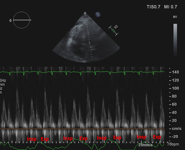
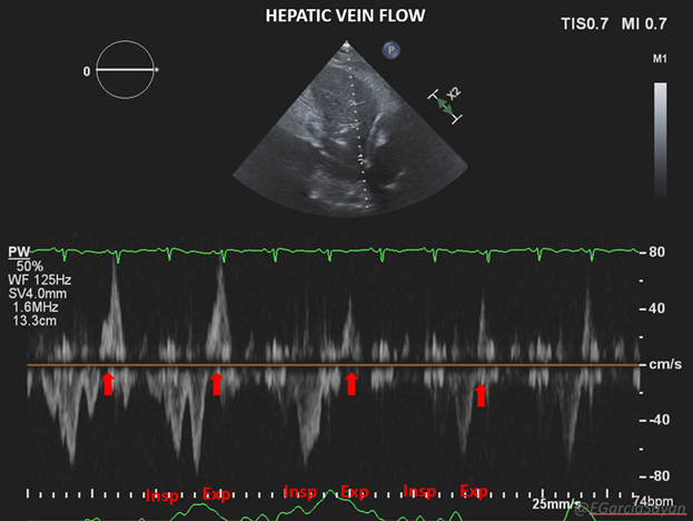
5/ PWD of the hepatic veins (HV) reveals expiratory diastolic flow reversal in both CP and CT. This is due to LV interdependence and septum shifting to the R in expiration, not allowing RV to fully expand in diastole. Demonstrated here (respirometer tracing not great, sorry...).
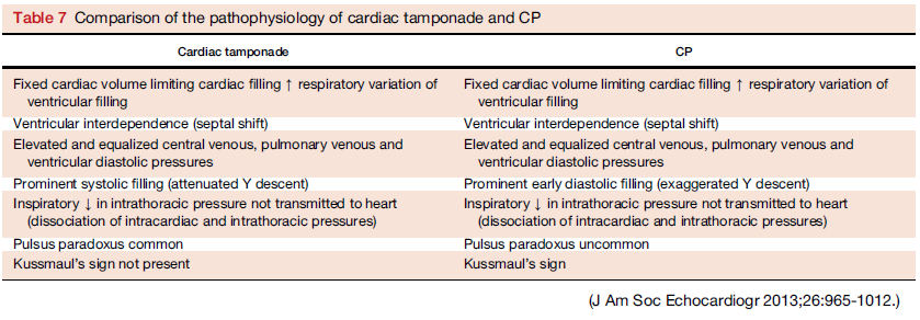
6/ So how do we differentiate CP from CT? First thing is of course the presence of significant pericardial effusion, clinical picture (pulsus paradoxus vs Kussmaul& #39;s sign), and R chamber collapse (for RA >30% of cardiac cycle). This case has an effusion but no definite collapse.
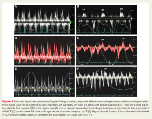
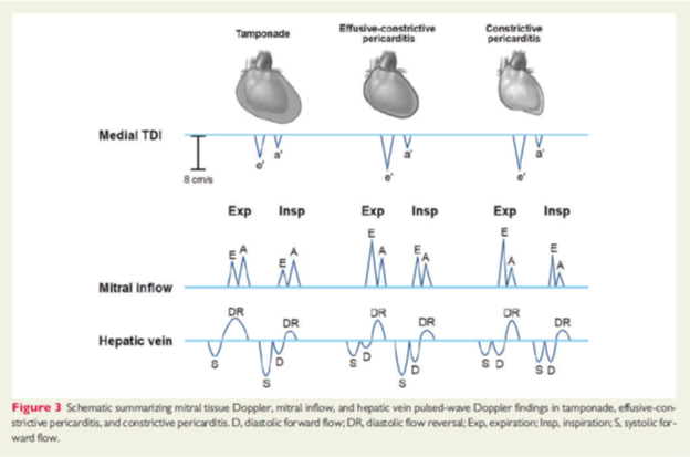
7/ Mitral inflow & HV flow can help differentiate CT from pure CP and effusive-constrictive pericarditis (ECP). All with insp reversal:
 https://abs.twimg.com/emoji/v2/... draggable="false" alt="🔶" title="Große orangene Raute" aria-label="Emoji: Große orangene Raute">CT: lower E/A ratio,
  https://abs.twimg.com/emoji/v2/... draggable="false" alt="🚫" title=""Betreten verboten!"-Zeichen" aria-label="Emoji: "Betreten verboten!"-Zeichen">HV diast forward flow
 https://abs.twimg.com/emoji/v2/... draggable="false" alt="🔶" title="Große orangene Raute" aria-label="Emoji: Große orangene Raute">CP: E>A, prominent HV diast forward flow
 https://abs.twimg.com/emoji/v2/... draggable="false" alt="🔶" title="Große orangene Raute" aria-label="Emoji: Große orangene Raute">ECP: Intermediate (+ organized/echogenic effusion?)
8/ So though CT, CP and ECP all display inspiratory decrease in mitral inflow and diastolic flow reversal in hepatic veins, PWD patterns in mitral inflow and HV are not the same!
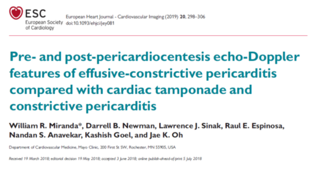
 https://abs.twimg.com/emoji/v2/... draggable="false" alt="👉" title="Rückhand Zeigefinger nach rechts" aria-label="Emoji: Rückhand Zeigefinger nach rechts">See excellent paper by @JaeKOh2 et al on this subject: https://academic.oup.com/ehjcimaging/article/20/3/298/5049591">https://academic.oup.com/ehjcimagi...
9/ End of " #tweetorial. Subtle cases of constrictive pericarditis can often be missed if not paying attention to these details. And differentiation of cardiac tamponade and constrictive physiology in patients with pericardial effusion is not always easy! I hope this was helpful.

 Read on Twitter
 There is constriction, but specifically effusive-constrictive pericarditis.https://abs.twimg.com/emoji/v2/... draggable="false" alt="👉" title="Rückhand Zeigefinger nach rechts" aria-label="Emoji: Rückhand Zeigefinger nach rechts"> #tweetorial here: https://abs.twimg.com/emoji/v2/... draggable="false" alt="🔶" title="Große orangene Raute" aria-label="Emoji: Große orangene Raute">To understand #EchoFirst findings in pericardial disease, must go back to basicshttps://abs.twimg.com/emoji/v2/... draggable="false" alt="🔶" title="Große orangene Raute" aria-label="Emoji: Große orangene Raute">Remember that superior portion of LA and pulmonary veins are extra-pericardial structures." title="1/ https://abs.twimg.com/emoji/v2/... draggable="false" alt="💡" title="Elektrische Glühbirne" aria-label="Emoji: Elektrische Glühbirne">There is constriction, but specifically effusive-constrictive pericarditis.https://abs.twimg.com/emoji/v2/... draggable="false" alt="👉" title="Rückhand Zeigefinger nach rechts" aria-label="Emoji: Rückhand Zeigefinger nach rechts"> #tweetorial here: https://abs.twimg.com/emoji/v2/... draggable="false" alt="🔶" title="Große orangene Raute" aria-label="Emoji: Große orangene Raute">To understand #EchoFirst findings in pericardial disease, must go back to basicshttps://abs.twimg.com/emoji/v2/... draggable="false" alt="🔶" title="Große orangene Raute" aria-label="Emoji: Große orangene Raute">Remember that superior portion of LA and pulmonary veins are extra-pericardial structures.">
 There is constriction, but specifically effusive-constrictive pericarditis.https://abs.twimg.com/emoji/v2/... draggable="false" alt="👉" title="Rückhand Zeigefinger nach rechts" aria-label="Emoji: Rückhand Zeigefinger nach rechts"> #tweetorial here: https://abs.twimg.com/emoji/v2/... draggable="false" alt="🔶" title="Große orangene Raute" aria-label="Emoji: Große orangene Raute">To understand #EchoFirst findings in pericardial disease, must go back to basicshttps://abs.twimg.com/emoji/v2/... draggable="false" alt="🔶" title="Große orangene Raute" aria-label="Emoji: Große orangene Raute">Remember that superior portion of LA and pulmonary veins are extra-pericardial structures." title="1/ https://abs.twimg.com/emoji/v2/... draggable="false" alt="💡" title="Elektrische Glühbirne" aria-label="Emoji: Elektrische Glühbirne">There is constriction, but specifically effusive-constrictive pericarditis.https://abs.twimg.com/emoji/v2/... draggable="false" alt="👉" title="Rückhand Zeigefinger nach rechts" aria-label="Emoji: Rückhand Zeigefinger nach rechts"> #tweetorial here: https://abs.twimg.com/emoji/v2/... draggable="false" alt="🔶" title="Große orangene Raute" aria-label="Emoji: Große orangene Raute">To understand #EchoFirst findings in pericardial disease, must go back to basicshttps://abs.twimg.com/emoji/v2/... draggable="false" alt="🔶" title="Große orangene Raute" aria-label="Emoji: Große orangene Raute">Remember that superior portion of LA and pulmonary veins are extra-pericardial structures.">
 of intra-thoracic pressure & in PVeins is not transmitted to LA or LV. This dissociation leads tohttps://abs.twimg.com/emoji/v2/... draggable="false" alt="⬇️" title="Pfeil nach unten" aria-label="Emoji: Pfeil nach unten">PVein-LA gradient and https://abs.twimg.com/emoji/v2/... draggable="false" alt="⬇️" title="Pfeil nach unten" aria-label="Emoji: Pfeil nach unten">LV filling, demonstrated by an inspiratory https://abs.twimg.com/emoji/v2/... draggable="false" alt="⬇️" title="Pfeil nach unten" aria-label="Emoji: Pfeil nach unten">in mitral inflow velocity by >25%." title="2/ In constrictive pericarditis (CP) or cardiac tamponade (CT), inspiratoryhttps://abs.twimg.com/emoji/v2/... draggable="false" alt="⬇️" title="Pfeil nach unten" aria-label="Emoji: Pfeil nach unten">of intra-thoracic pressure & in PVeins is not transmitted to LA or LV. This dissociation leads tohttps://abs.twimg.com/emoji/v2/... draggable="false" alt="⬇️" title="Pfeil nach unten" aria-label="Emoji: Pfeil nach unten">PVein-LA gradient and https://abs.twimg.com/emoji/v2/... draggable="false" alt="⬇️" title="Pfeil nach unten" aria-label="Emoji: Pfeil nach unten">LV filling, demonstrated by an inspiratory https://abs.twimg.com/emoji/v2/... draggable="false" alt="⬇️" title="Pfeil nach unten" aria-label="Emoji: Pfeil nach unten">in mitral inflow velocity by >25%." class="img-responsive" style="max-width:100%;"/>
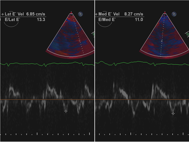 e& #39; and https://abs.twimg.com/emoji/v2/... draggable="false" alt="⬆️" title="Pfeil nach oben" aria-label="Emoji: Pfeil nach oben"> E/e& #39;) but in constriction e& #39; is preserved and often higher in the septal annulus rather than lateral which can be tethered by the thickened pericardium ("annulus reversus" or "annulus paradoxus") as seen in our case." title="4/ In restriction, tissue Doppler is abnormal (https://abs.twimg.com/emoji/v2/... draggable="false" alt="⬇️" title="Pfeil nach unten" aria-label="Emoji: Pfeil nach unten">e& #39; and https://abs.twimg.com/emoji/v2/... draggable="false" alt="⬆️" title="Pfeil nach oben" aria-label="Emoji: Pfeil nach oben"> E/e& #39;) but in constriction e& #39; is preserved and often higher in the septal annulus rather than lateral which can be tethered by the thickened pericardium ("annulus reversus" or "annulus paradoxus") as seen in our case." class="img-responsive" style="max-width:100%;"/>
 See excellent paper by @JaeKOh2 et al on this subject: https://academic.oup.com/ehjcimagi..." title="8/ So though CT, CP and ECP all display inspiratory decrease in mitral inflow and diastolic flow reversal in hepatic veins, PWD patterns in mitral inflow and HV are not the same! https://abs.twimg.com/emoji/v2/... draggable="false" alt="👉" title="Rückhand Zeigefinger nach rechts" aria-label="Emoji: Rückhand Zeigefinger nach rechts">See excellent paper by @JaeKOh2 et al on this subject: https://academic.oup.com/ehjcimagi...">
 See excellent paper by @JaeKOh2 et al on this subject: https://academic.oup.com/ehjcimagi..." title="8/ So though CT, CP and ECP all display inspiratory decrease in mitral inflow and diastolic flow reversal in hepatic veins, PWD patterns in mitral inflow and HV are not the same! https://abs.twimg.com/emoji/v2/... draggable="false" alt="👉" title="Rückhand Zeigefinger nach rechts" aria-label="Emoji: Rückhand Zeigefinger nach rechts">See excellent paper by @JaeKOh2 et al on this subject: https://academic.oup.com/ehjcimagi...">
 See excellent paper by @JaeKOh2 et al on this subject: https://academic.oup.com/ehjcimagi..." title="8/ So though CT, CP and ECP all display inspiratory decrease in mitral inflow and diastolic flow reversal in hepatic veins, PWD patterns in mitral inflow and HV are not the same! https://abs.twimg.com/emoji/v2/... draggable="false" alt="👉" title="Rückhand Zeigefinger nach rechts" aria-label="Emoji: Rückhand Zeigefinger nach rechts">See excellent paper by @JaeKOh2 et al on this subject: https://academic.oup.com/ehjcimagi...">


