1/ #Nephrology #POCUS case study:
Dr. X is rounding on an ESRD pt who initially presented with dyspnea after missing a dialysis (HD) session; underwent dialysis in the hospital. Pt asymptomatic at the time of exam and lung #ultrasound revealed https://abs.twimg.com/emoji/v2/... draggable="false" alt="👇" title="Rückhand Zeigefinger nach unten" aria-label="Emoji: Rückhand Zeigefinger nach unten"> Further story in thread #MedEd
Dr. X is rounding on an ESRD pt who initially presented with dyspnea after missing a dialysis (HD) session; underwent dialysis in the hospital. Pt asymptomatic at the time of exam and lung #ultrasound revealed
2/ Based on the 2-zone lung #POCUS, Dr. X orders for another session of HD. Notably, pt says he is at his & #39;dry weight& #39; and HD nurse says they could only get 1.5L off during first session. Dr. X doesn& #39;t change his/her mind.
Info on various lung scan zones https://abs.twimg.com/emoji/v2/... draggable="false" alt="👇" title="Rückhand Zeigefinger nach unten" aria-label="Emoji: Rückhand Zeigefinger nach unten"> https://youtu.be/o9Syl0F3AuY ">https://youtu.be/o9Syl0F3A...
Info on various lung scan zones
3/ Patient becomes hypotensive during HD and only ~500cc fluid could be removed.
Why can& #39;t we get more fluid out of a hypervolemic patient? Dr. X is perplexed and decides to more #POCUS Here is the IVC
Why can& #39;t we get more fluid out of a hypervolemic patient? Dr. X is perplexed and decides to more #POCUS Here is the IVC
4/ Looks like the tank is empty....no wonder pt didn& #39;t tolerate ultrafiltration.
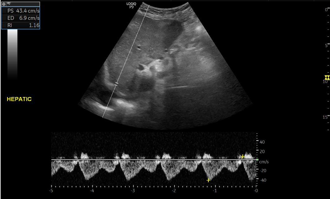
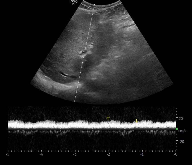
#VExUS is not needed if IVC is small but just to make sure, hepatic and portal vein Doppler obtained - look normal!
#VExUS is not needed if IVC is small but just to make sure, hepatic and portal vein Doppler obtained - look normal!
5/ Time to scan the lungs again....
This time Dr. X does 8-zone #POCUS scan and finds similar images.
This time Dr. X does 8-zone #POCUS scan and finds similar images.
6/ Why are the lungs still congested? patient is asymptomatic. May be heart is the culprit  https://abs.twimg.com/emoji/v2/... draggable="false" alt="🤔" title="Denkendes Gesicht" aria-label="Emoji: Denkendes Gesicht">
Pump function? diastolic dysfunction? bad mitral regurgitation?
Cardiac #POCUS performed. Not great windows but here is what Dr. X managed to get. Subcostal https://abs.twimg.com/emoji/v2/... draggable="false" alt="👇" title="Rückhand Zeigefinger nach unten" aria-label="Emoji: Rückhand Zeigefinger nach unten">
Pump function? diastolic dysfunction? bad mitral regurgitation?
Cardiac #POCUS performed. Not great windows but here is what Dr. X managed to get. Subcostal
7/ No pericardial effusion, LV systolic function grossly seems OK.
Little bit of maneuvering shows some aortic valve calcification. Aortic stenosis? gradient not measured but seems to be opening, LV doesn& #39;t look obviously big/thick. Pt is 60+, kidney disease.
Little bit of maneuvering shows some aortic valve calcification. Aortic stenosis? gradient not measured but seems to be opening, LV doesn& #39;t look obviously big/thick. Pt is 60+, kidney disease.
8/ Moving on to apical window #POCUS. Grey scale looks OK, may be some mitral annular calcification (note shadowing), which is also not unexpected in patients with kidney disease.
9/ Color: May not be optimal but doesn& #39;t look like there is horrible mitral regurgitation. May be a little aortic regurgitation  https://abs.twimg.com/emoji/v2/... draggable="false" alt="🤔" title="Denkendes Gesicht" aria-label="Emoji: Denkendes Gesicht">
10/ Another color
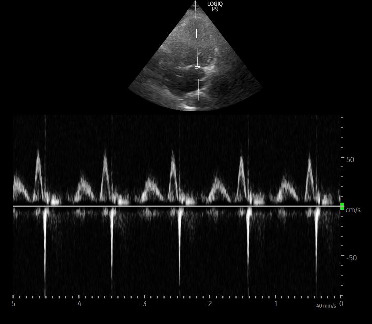
11/ Mitral inflow Doppler #POCUS
Impaired relaxation (A>E) but not unexpected in older people. Shouldn& #39;t fill the lung with B-lines. Tissue Doppler not performed.
Need a refresher on diastology? Refer to @Pocus101 & #39;s guide https://www.pocus101.com/how-to-measure-and-grade-diastolic-dysfunction-using-echocardiography/">https://www.pocus101.com/how-to-me...
12/ Lung #POCUS performed again. Dr. X noted something unusual about the pleural line. Takes a closer look with linear probe (high resolution - lower depth)
Left lung images https://abs.twimg.com/emoji/v2/... draggable="false" alt="👇" title="Rückhand Zeigefinger nach unten" aria-label="Emoji: Rückhand Zeigefinger nach unten">
Left lung images
13/ Right lung #POCUS
Irregular pleural line suggestive of underlying lung disease (COVID negative, no symptoms). A CT chest was ordered.
Irregular pleural line suggestive of underlying lung disease (COVID negative, no symptoms). A CT chest was ordered.
14/ CT demonstrated interlobular septal thickening more pronounced at bases and linear scarring more pronounced in the upper lobes. Pt chronic smoker.
Verdict: B-lines on lung #POCUS were likely not due to water. Pt referred to Pulmonologist.
Verdict: B-lines on lung #POCUS were likely not due to water. Pt referred to Pulmonologist.
15/ B-lines can be seen in lung fibrosis, contusion, pneumonia etc. in addition to congestion/edema.
In these cases, pleural line is irregular, show subpleural consolidations, may be thickened and some areas are spared.
Diffuse involvement as in this Pt can create confusion.
In these cases, pleural line is irregular, show subpleural consolidations, may be thickened and some areas are spared.
Diffuse involvement as in this Pt can create confusion.
16/ What if this patient really has pulmonary edema? How can lung #POCUS help in the outpatient #dialysis unit?
Any input @kyliebaker888 @siddharth_dugar @msiuba https://abs.twimg.com/emoji/v2/... draggable="false" alt="🧐" title="Gesicht mit Monokel" aria-label="Emoji: Gesicht mit Monokel">
Any input @kyliebaker888 @siddharth_dugar @msiuba

 Read on Twitter


