#NeurologyMorningReport Case 43 #MedTwitter Updates & Answers posted later today. Asking your help #MedEd #neurology #neurologyresident #neurologist #medstudent #NeurologyProud #IamaNeurologist Join me in educating. Share your questions and knowledge.
1/
1/
65 yo man h/o diabetes p/w progressive dysphagia, diplopia, &dysarthria x3m.
Thinking fast- show your biases- what& #39;s 1st diagnosis that comes to mind given this history? If you want to be anonymous respond via polleverywhere (no-spaces-between-words) -word cloud later today
2/
Thinking fast- show your biases- what& #39;s 1st diagnosis that comes to mind given this history? If you want to be anonymous respond via polleverywhere (no-spaces-between-words) -word cloud later today
2/
Initially, he described fluctuating symptoms and as part of w/u for MG, had CT scan of the chest.
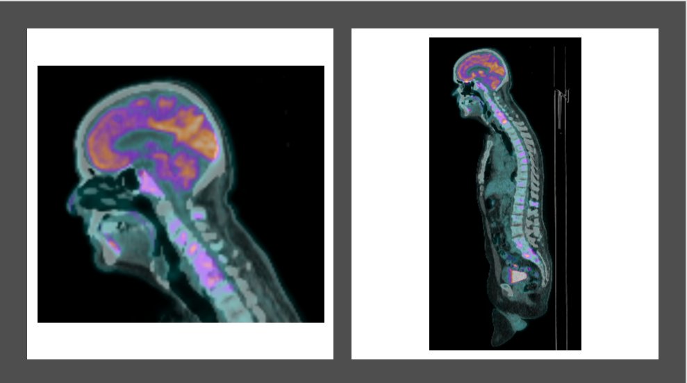
CT showed multiple osteolytic & blastic lesions in vertebral bodies and ribs.
More history revealed weight loss and fatigue over months.
PE: representative video and images
3/
CT showed multiple osteolytic & blastic lesions in vertebral bodies and ribs.
More history revealed weight loss and fatigue over months.
PE: representative video and images
3/
Abnormalities on exam shown in video and below drawing and image (from another pt).
Mental status and neuro exam below the neck were normal.
4/
Mental status and neuro exam below the neck were normal.
4/
Where do you localize the lesion?
Great tweetorial by @AaronLBerkowitz on brainstem anatomy and cranial nerves https://threadreaderapp.com/thread/1265051075431690240.html
Please">https://threadreaderapp.com/thread/12... add other resources you recommend for learning about significance of this patient& #39;s findings.
5/
Great tweetorial by @AaronLBerkowitz on brainstem anatomy and cranial nerves https://threadreaderapp.com/thread/1265051075431690240.html
Please">https://threadreaderapp.com/thread/12... add other resources you recommend for learning about significance of this patient& #39;s findings.
5/
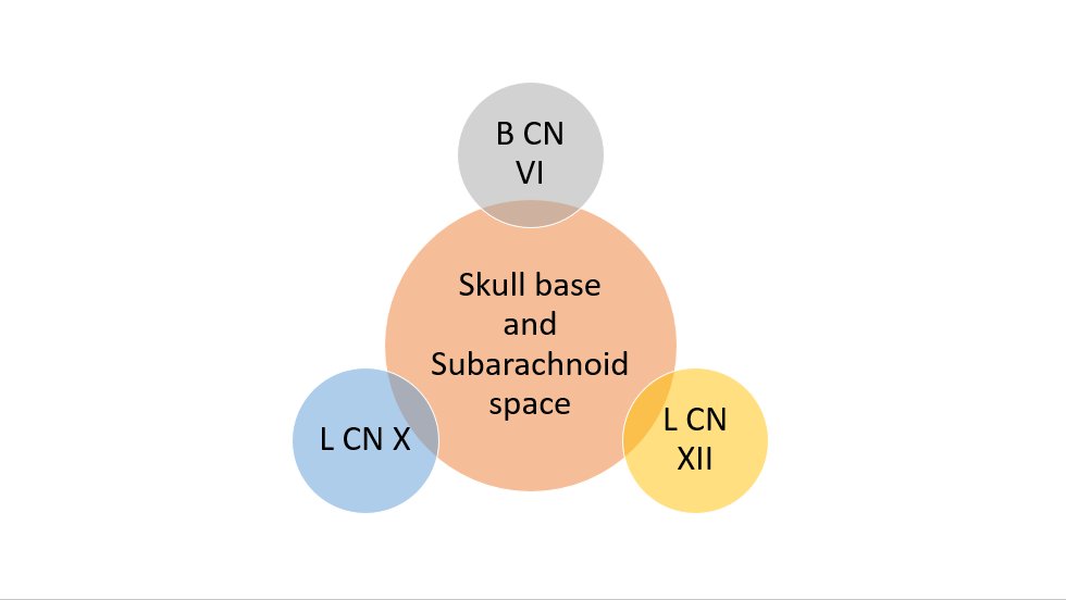
Video shows bilateral abducens palsies (B CN VI)
Drawing shows L palate weakness (L CN X)
Image shows L tongue atrophy (L CN XII). The involvement of the eye movements means that it cannot only localize to the medulla.
6/
Drawing shows L palate weakness (L CN X)
Image shows L tongue atrophy (L CN XII). The involvement of the eye movements means that it cannot only localize to the medulla.
6/
PSA makedly elevated. Bx shows:
Dx: #ProstateCancer with #SkullBaseMetastases #MultipleCranialNeuropathies #GodtfredsenSyndrome
Other examples:
https://www.sciencedirect.com/science/article/abs/pii/S152918390600697X
https://www.sciencedirect.com/science/a... href=" https://acsjournals.onlinelibrary.wiley.com/doi/full/10.1002/cncr.20553">https://acsjournals.onlinelibrary.wiley.com/doi/full/... https://www.ncbi.nlm.nih.gov/pmc/articles/PMC3212593/">https://www.ncbi.nlm.nih.gov/pmc/artic...
Dx: #ProstateCancer with #SkullBaseMetastases #MultipleCranialNeuropathies #GodtfredsenSyndrome
Other examples:
https://www.sciencedirect.com/science/article/abs/pii/S152918390600697X

 Read on Twitter


