Stop KILLING Your Patients with Fluid! Learn how to avoid Fluid Overload with Venous Congestion Evaluation using #POCUS
New BLOG POST on the #VExUS Protocol! Step By Step Guide, Downloadable PDF Pocket Guide, & Calculator
 https://abs.twimg.com/emoji/v2/... draggable="false" alt="👉" title="Right pointing backhand index" aria-label="Emoji: Right pointing backhand index">
 https://abs.twimg.com/emoji/v2/... draggable="false" alt="🔗" title="Link symbol" aria-label="Emoji: Link symbol"> https://pocus101.com/vexus
https://pocus101.com/vexus&quo... href="https://twtext.com//hashtag/medtweetorial"> #medtweetorial https://abs.twimg.com/emoji/v2/... draggable="false" alt="👇" title="Down pointing backhand index" aria-label="Emoji: Down pointing backhand index">(1/10)
New BLOG POST on the #VExUS Protocol! Step By Step Guide, Downloadable PDF Pocket Guide, & Calculator
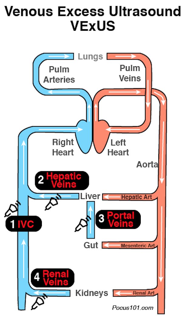
(2/10) It’s not just about the IVC it’s about what is affected beyond the IVC. Don’t forget the Liver, Gut, and Kidneys.
Venous Congestion in these organs can lead to significant comorbidity. Download the PDF: https://abs.twimg.com/emoji/v2/... draggable="false" alt="👉" title="Right pointing backhand index" aria-label="Emoji: Right pointing backhand index">
 https://abs.twimg.com/emoji/v2/... draggable="false" alt="🔗" title="Link symbol" aria-label="Emoji: Link symbol"> https://pocus101.com/vexus ">https://pocus101.com/vexus&quo...
Venous Congestion in these organs can lead to significant comorbidity. Download the PDF:
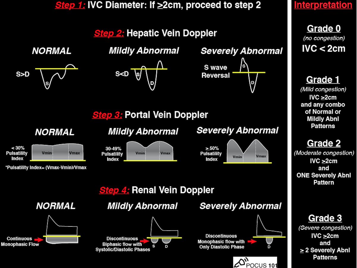
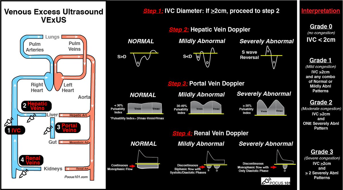
(3/10) Step 1: Look at the IVC if < 2cm then Grade 0, If > 2cm proceed to the next steps.  https://abs.twimg.com/emoji/v2/... draggable="false" alt="🔗" title="Link symbol" aria-label="Emoji: Link symbol"> https://pocus101.com/vexus ">https://pocus101.com/vexus&quo...
(4/10) Step 2: Evaluate Hepatic Venous Doppler:
 https://abs.twimg.com/emoji/v2/... draggable="false" alt="🔗" title="Link symbol" aria-label="Emoji: Link symbol"> https://pocus101.com/vexus
1.">https://pocus101.com/vexus&quo... Get Image of IVC and hepatic veins.
2. Place color flow Doppler over the hepatic veins. (Should see BLUE-away)
3. Place your Pulse Wave Doppler gate on a hepatic vein
4. Initiate Pulse wave Doppler
1.">https://pocus101.com/vexus&quo... Get Image of IVC and hepatic veins.
2. Place color flow Doppler over the hepatic veins. (Should see BLUE-away)
3. Place your Pulse Wave Doppler gate on a hepatic vein
4. Initiate Pulse wave Doppler
(5/10) Interpret Hepatic Venous Doppler Tracing.  https://abs.twimg.com/emoji/v2/... draggable="false" alt="🔗" title="Link symbol" aria-label="Emoji: Link symbol"> https://pocus101.com/vexus ">https://pocus101.com/vexus&quo...
(6/10) Step 3: Evaluate Portal Venous Doppler:
 https://abs.twimg.com/emoji/v2/... draggable="false" alt="🔗" title="Link symbol" aria-label="Emoji: Link symbol"> https://pocus101.com/vexus
1.">https://pocus101.com/vexus&quo... Get 2D image of the Right Portal Vein
2. Place color flow Doppler over Portal Vein (Should See RED-toward)
3. Place your pulse wave Doppler gate on the Portal Vein
4. Initiate Pulse wave Doppler
1.">https://pocus101.com/vexus&quo... Get 2D image of the Right Portal Vein
2. Place color flow Doppler over Portal Vein (Should See RED-toward)
3. Place your pulse wave Doppler gate on the Portal Vein
4. Initiate Pulse wave Doppler
(7/9) Interpret Portal Venous Doppler Tracing.  https://abs.twimg.com/emoji/v2/... draggable="false" alt="🔗" title="Link symbol" aria-label="Emoji: Link symbol"> https://pocus101.com/vexus ">https://pocus101.com/vexus&quo...
(8/10) Step 2: Evaluate Intrarenal Venous Doppler:
 https://abs.twimg.com/emoji/v2/... draggable="false" alt="🔗" title="Link symbol" aria-label="Emoji: Link symbol"> https://pocus101.com/vexus
1.">https://pocus101.com/vexus&quo... Get a 2-D image of the Kidney
2. Place color flow Doppler over Kidney.
3. Place your pulse wave Doppler gate on the interlobar vessels
4. Initiate Pulse wave Doppler
Clip https://abs.twimg.com/emoji/v2/... draggable="false" alt="👇" title="Down pointing backhand index" aria-label="Emoji: Down pointing backhand index"> https://www.youtube.com/watch?v=0I9fi2S0nKw&feature=emb_title">https://www.youtube.com/watch...
1.">https://pocus101.com/vexus&quo... Get a 2-D image of the Kidney
2. Place color flow Doppler over Kidney.
3. Place your pulse wave Doppler gate on the interlobar vessels
4. Initiate Pulse wave Doppler
Clip
(9/10) Interpret Renal Venous Doppler Tracing.  https://abs.twimg.com/emoji/v2/... draggable="false" alt="🔗" title="Link symbol" aria-label="Emoji: Link symbol"> https://pocus101.com/vexus ">https://pocus101.com/vexus&quo...
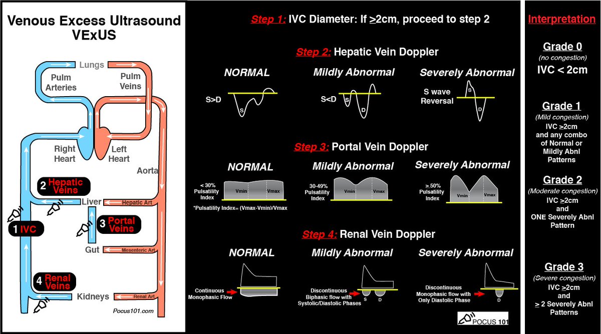
(9/10) VExUS Severity Grading:
Download PDF! https://abs.twimg.com/emoji/v2/... draggable="false" alt="👉" title="Right pointing backhand index" aria-label="Emoji: Right pointing backhand index">
 https://abs.twimg.com/emoji/v2/... draggable="false" alt="🔗" title="Link symbol" aria-label="Emoji: Link symbol"> https://pocus101.com/vexus
Grade">https://pocus101.com/vexus&quo... 0: IVC <2cm = NO Congestion
Grade 1: IVC >=2cm with any combo of Normal or Mildly Abnl Patterns
Grade 2: IVC >=2cm and ONE severely Abnl Pattern
Grade 3: IVC >=2cm and >2 Severely Abnl Patterns
Download PDF!
Grade">https://pocus101.com/vexus&quo... 0: IVC <2cm = NO Congestion
Grade 1: IVC >=2cm with any combo of Normal or Mildly Abnl Patterns
Grade 2: IVC >=2cm and ONE severely Abnl Pattern
Grade 3: IVC >=2cm and >2 Severely Abnl Patterns
Special thanks to my colleague @khaycock2 for reviewing this material for me. And of course the #VExUS Pioneers: @ThinkingCC @khaycock2 @WBeaubien @EMNerd_ And the #VExUS superusers! @ArgaizR @NephroP @Wilkinsonjonny @Thind888 @load_dependent. Learning from all of you!  https://abs.twimg.com/emoji/v2/... draggable="false" alt="🙏" title="Folded hands" aria-label="Emoji: Folded hands">
 https://abs.twimg.com/emoji/v2/... draggable="false" alt="🙏" title="Folded hands" aria-label="Emoji: Folded hands">

 Read on Twitter
 https://abs.twimg.com/emoji/v2/... draggable="false" alt="🔗" title="Link symbol" aria-label="Emoji: Link symbol"> https://pocus101.com/vexus&quo... href="https://twtext.com//hashtag/medtweetorial"> #medtweetorialhttps://abs.twimg.com/emoji/v2/... draggable="false" alt="👇" title="Down pointing backhand index" aria-label="Emoji: Down pointing backhand index">(1/10)" title="Stop KILLING Your Patients with Fluid! Learn how to avoid Fluid Overload with Venous Congestion Evaluation using #POCUSNew BLOG POST on the #VExUS Protocol! Step By Step Guide, Downloadable PDF Pocket Guide, & Calculator https://abs.twimg.com/emoji/v2/... draggable="false" alt="👉" title="Right pointing backhand index" aria-label="Emoji: Right pointing backhand index">https://abs.twimg.com/emoji/v2/... draggable="false" alt="🔗" title="Link symbol" aria-label="Emoji: Link symbol"> https://pocus101.com/vexus&quo... href="https://twtext.com//hashtag/medtweetorial"> #medtweetorialhttps://abs.twimg.com/emoji/v2/... draggable="false" alt="👇" title="Down pointing backhand index" aria-label="Emoji: Down pointing backhand index">(1/10)">
 https://abs.twimg.com/emoji/v2/... draggable="false" alt="🔗" title="Link symbol" aria-label="Emoji: Link symbol"> https://pocus101.com/vexus&quo... href="https://twtext.com//hashtag/medtweetorial"> #medtweetorialhttps://abs.twimg.com/emoji/v2/... draggable="false" alt="👇" title="Down pointing backhand index" aria-label="Emoji: Down pointing backhand index">(1/10)" title="Stop KILLING Your Patients with Fluid! Learn how to avoid Fluid Overload with Venous Congestion Evaluation using #POCUSNew BLOG POST on the #VExUS Protocol! Step By Step Guide, Downloadable PDF Pocket Guide, & Calculator https://abs.twimg.com/emoji/v2/... draggable="false" alt="👉" title="Right pointing backhand index" aria-label="Emoji: Right pointing backhand index">https://abs.twimg.com/emoji/v2/... draggable="false" alt="🔗" title="Link symbol" aria-label="Emoji: Link symbol"> https://pocus101.com/vexus&quo... href="https://twtext.com//hashtag/medtweetorial"> #medtweetorialhttps://abs.twimg.com/emoji/v2/... draggable="false" alt="👇" title="Down pointing backhand index" aria-label="Emoji: Down pointing backhand index">(1/10)">
 https://abs.twimg.com/emoji/v2/... draggable="false" alt="🔗" title="Link symbol" aria-label="Emoji: Link symbol"> https://pocus101.com/vexus&quo... href="https://twtext.com//hashtag/medtweetorial"> #medtweetorialhttps://abs.twimg.com/emoji/v2/... draggable="false" alt="👇" title="Down pointing backhand index" aria-label="Emoji: Down pointing backhand index">(1/10)" title="Stop KILLING Your Patients with Fluid! Learn how to avoid Fluid Overload with Venous Congestion Evaluation using #POCUSNew BLOG POST on the #VExUS Protocol! Step By Step Guide, Downloadable PDF Pocket Guide, & Calculator https://abs.twimg.com/emoji/v2/... draggable="false" alt="👉" title="Right pointing backhand index" aria-label="Emoji: Right pointing backhand index">https://abs.twimg.com/emoji/v2/... draggable="false" alt="🔗" title="Link symbol" aria-label="Emoji: Link symbol"> https://pocus101.com/vexus&quo... href="https://twtext.com//hashtag/medtweetorial"> #medtweetorialhttps://abs.twimg.com/emoji/v2/... draggable="false" alt="👇" title="Down pointing backhand index" aria-label="Emoji: Down pointing backhand index">(1/10)">
 https://abs.twimg.com/emoji/v2/... draggable="false" alt="🔗" title="Link symbol" aria-label="Emoji: Link symbol"> https://pocus101.com/vexus&quo..." title="(2/10) It’s not just about the IVC it’s about what is affected beyond the IVC. Don’t forget the Liver, Gut, and Kidneys.Venous Congestion in these organs can lead to significant comorbidity. Download the PDF: https://abs.twimg.com/emoji/v2/... draggable="false" alt="👉" title="Right pointing backhand index" aria-label="Emoji: Right pointing backhand index">https://abs.twimg.com/emoji/v2/... draggable="false" alt="🔗" title="Link symbol" aria-label="Emoji: Link symbol"> https://pocus101.com/vexus&quo..." class="img-responsive" style="max-width:100%;"/>
 https://pocus101.com/vexus&quo... Get Image of IVC and hepatic veins.2. Place color flow Doppler over the hepatic veins. (Should see BLUE-away)3. Place your Pulse Wave Doppler gate on a hepatic vein4. Initiate Pulse wave Doppler" title="(4/10) Step 2: Evaluate Hepatic Venous Doppler:https://abs.twimg.com/emoji/v2/... draggable="false" alt="🔗" title="Link symbol" aria-label="Emoji: Link symbol"> https://pocus101.com/vexus&quo... Get Image of IVC and hepatic veins.2. Place color flow Doppler over the hepatic veins. (Should see BLUE-away)3. Place your Pulse Wave Doppler gate on a hepatic vein4. Initiate Pulse wave Doppler" class="img-responsive" style="max-width:100%;"/>
 https://pocus101.com/vexus&quo..." title="(5/10) Interpret Hepatic Venous Doppler Tracing. https://abs.twimg.com/emoji/v2/... draggable="false" alt="🔗" title="Link symbol" aria-label="Emoji: Link symbol"> https://pocus101.com/vexus&quo..." class="img-responsive" style="max-width:100%;"/>
 https://pocus101.com/vexus&quo... Get 2D image of the Right Portal Vein2. Place color flow Doppler over Portal Vein (Should See RED-toward)3. Place your pulse wave Doppler gate on the Portal Vein4. Initiate Pulse wave Doppler" title="(6/10) Step 3: Evaluate Portal Venous Doppler:https://abs.twimg.com/emoji/v2/... draggable="false" alt="🔗" title="Link symbol" aria-label="Emoji: Link symbol"> https://pocus101.com/vexus&quo... Get 2D image of the Right Portal Vein2. Place color flow Doppler over Portal Vein (Should See RED-toward)3. Place your pulse wave Doppler gate on the Portal Vein4. Initiate Pulse wave Doppler" class="img-responsive" style="max-width:100%;"/>
 https://pocus101.com/vexus&quo..." title="(7/9) Interpret Portal Venous Doppler Tracing. https://abs.twimg.com/emoji/v2/... draggable="false" alt="🔗" title="Link symbol" aria-label="Emoji: Link symbol"> https://pocus101.com/vexus&quo..." class="img-responsive" style="max-width:100%;"/>
 https://pocus101.com/vexus&quo..." title="(9/10) Interpret Renal Venous Doppler Tracing. https://abs.twimg.com/emoji/v2/... draggable="false" alt="🔗" title="Link symbol" aria-label="Emoji: Link symbol"> https://pocus101.com/vexus&quo..." class="img-responsive" style="max-width:100%;"/>
 https://abs.twimg.com/emoji/v2/... draggable="false" alt="🔗" title="Link symbol" aria-label="Emoji: Link symbol"> https://pocus101.com/vexus&quo... 0: IVC <2cm = NO CongestionGrade 1: IVC >=2cm with any combo of Normal or Mildly Abnl PatternsGrade 2: IVC >=2cm and ONE severely Abnl PatternGrade 3: IVC >=2cm and >2 Severely Abnl Patterns" title="(9/10) VExUS Severity Grading:Download PDF! https://abs.twimg.com/emoji/v2/... draggable="false" alt="👉" title="Right pointing backhand index" aria-label="Emoji: Right pointing backhand index">https://abs.twimg.com/emoji/v2/... draggable="false" alt="🔗" title="Link symbol" aria-label="Emoji: Link symbol"> https://pocus101.com/vexus&quo... 0: IVC <2cm = NO CongestionGrade 1: IVC >=2cm with any combo of Normal or Mildly Abnl PatternsGrade 2: IVC >=2cm and ONE severely Abnl PatternGrade 3: IVC >=2cm and >2 Severely Abnl Patterns" class="img-responsive" style="max-width:100%;"/>


