#MorphologyMonday
1. Whenever we look at blood films everything appears 2D, but red cells are in fact 3D. What do they actually look like? Be ready to be mind blown. #itsathread https://abs.twimg.com/emoji/v2/... draggable="false" alt="👇🏾" title="Down pointing backhand index (medium dark skin tone)" aria-label="Emoji: Down pointing backhand index (medium dark skin tone)">
#underthescope #biomedicalscientist #haematology #bloodfilm #redcells #morphology
1. Whenever we look at blood films everything appears 2D, but red cells are in fact 3D. What do they actually look like? Be ready to be mind blown. #itsathread
#underthescope #biomedicalscientist #haematology #bloodfilm #redcells #morphology
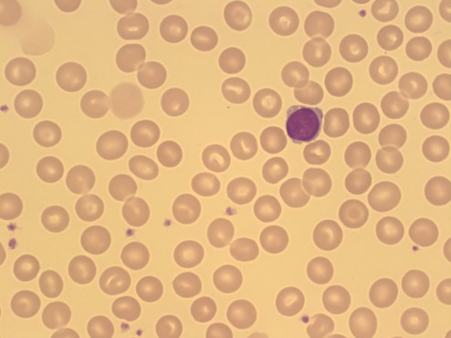
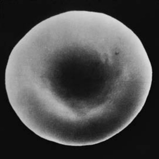
2. Normal red blood cells (erythrocytes) are disciform / biconcave in shape and appear on the blood film with a lighter area in the middle known as & #39;central pallor& #39;. This usually spans 1/3 of the diameter of the cell.
#MorphologyMonday
#MorphologyMonday
3. Reticulocytes are usually larger than normal red blood cells and they appear blueish due to staining of residual ribosomal RNA left after the nucleus is removed. Only the most immature reticulocytes appear polychromatic (blueish).
#MorphologyMonday
#MorphologyMonday
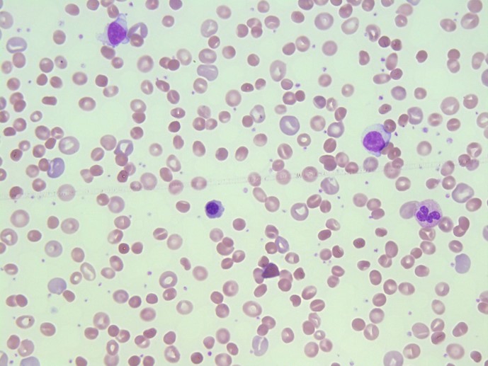
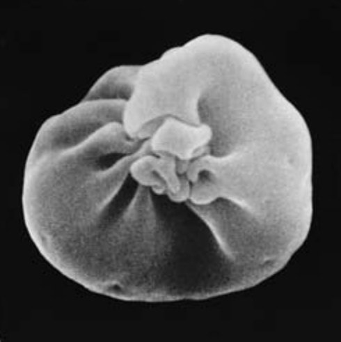
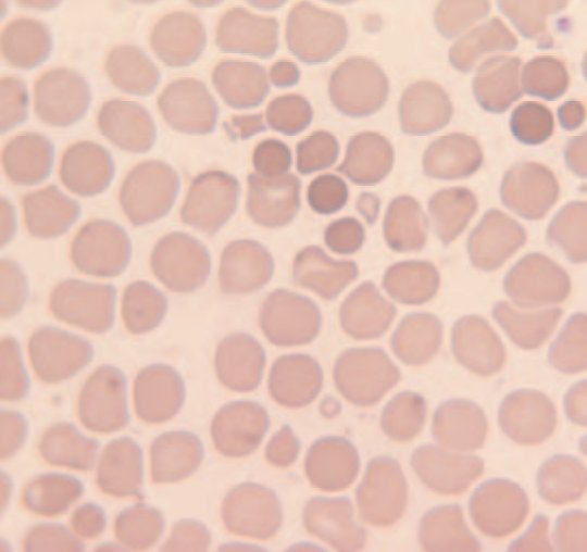
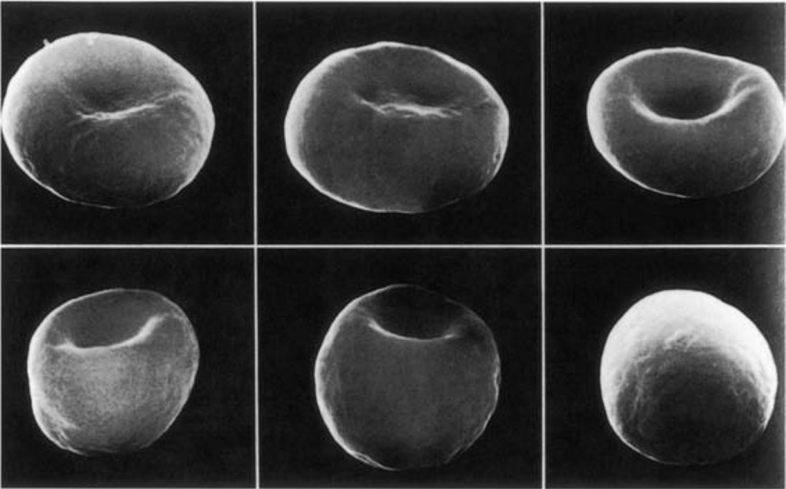
4. Spherocytes are as the name suggests, spherical rather than being disc shaped. They are cells that have lost membrane without equivalent loss of cytosol and appear dense / dark on the film. The SEM image shows stages from discocyte to spherocyte.
#MorphologyMonday
#MorphologyMonday
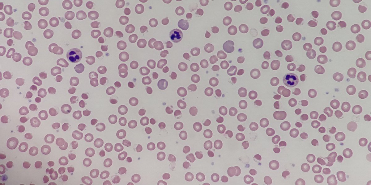
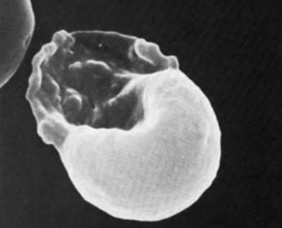
5. Hemighost cells occur when haemoglobin is damaged, usually where there is oxidative stress. This is indicative of G6PD deficiency. The haemoglobin is dense and contracts to one pole of the cell leaving an & #39;empty& #39; area surrounded by membrane.
#MorphologyMonday
#MorphologyMonday
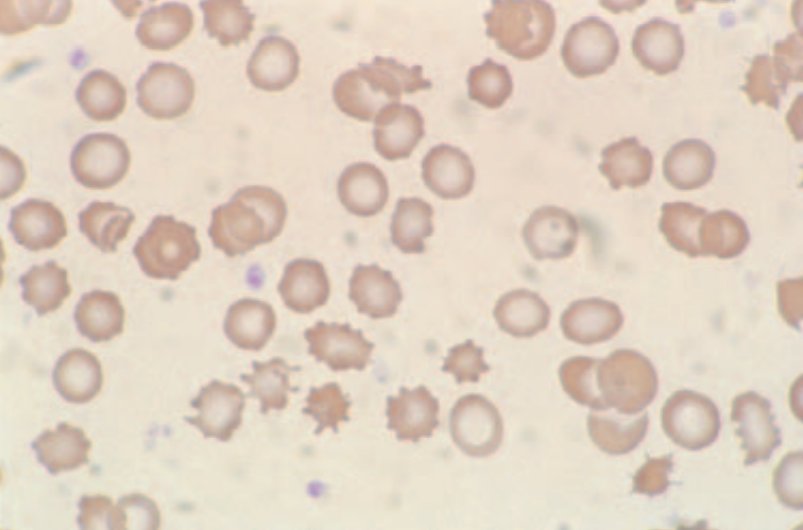
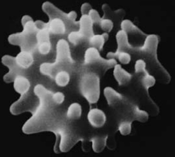
6. Echinocytes are red cells that have lost their distinct disc shape and are covered with 10–30 short blunt regular spicules. There are various reasons why they form including HUS, PK deficiency, storage artefact, liver disease and following burns for eg.
#MorphologyMonday
#MorphologyMonday
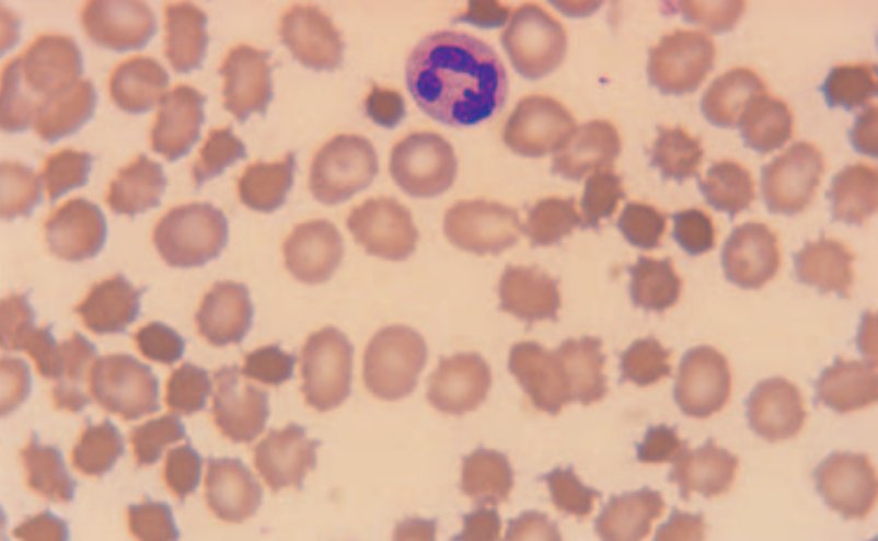
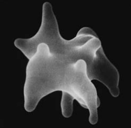
7. Acanthocytes are red cells bearing between 2-20 spicules that are of unequal length and distributed irregularly over the red cell surface. They& #39;re seen in abetalipoproteinaemia or liver disease, where cholesterol : phospholipid in red cells increases.
#MorphologyMonday
#MorphologyMonday
8. Keratocytes or & #39;horned cells& #39; usually have 2 / up to 4/6 spicules after opposing membranes fuse forming a pseudovacuole which then ruptures. These are seen in mechanical damage of the cells, MAHA, DIC, renal disease and after removal of Heinz bodies.
#MorphologyMonday
#MorphologyMonday
9. Target cells appear #underthescope as having an area of increased staining in the middle giving a  https://abs.twimg.com/emoji/v2/... draggable="false" alt="🎯" title="Direct hit" aria-label="Emoji: Direct hit"> like appearance. They are thinner than normal cells. In vivo they appear bell shaped but flatten on spreading giving the characteristic target shape.
#MorphologyMonday
#MorphologyMonday
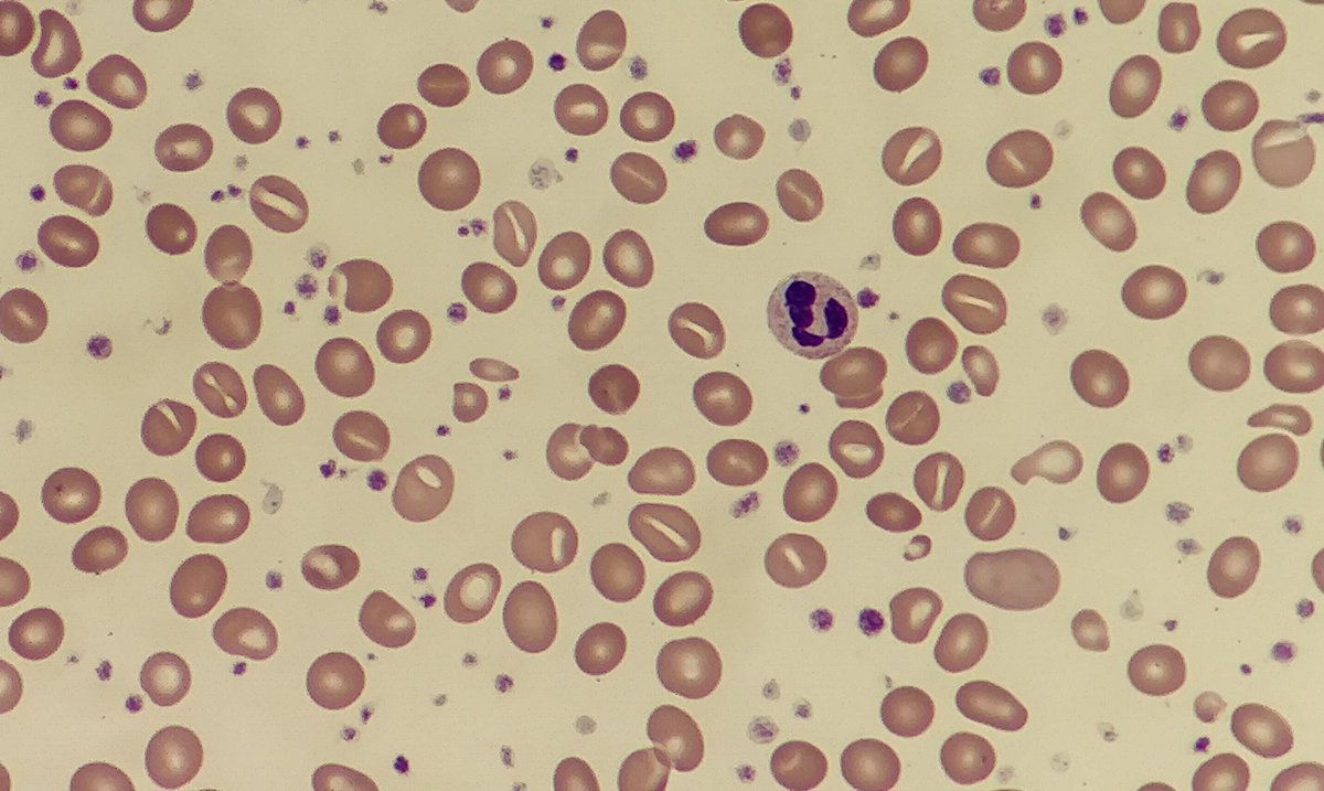
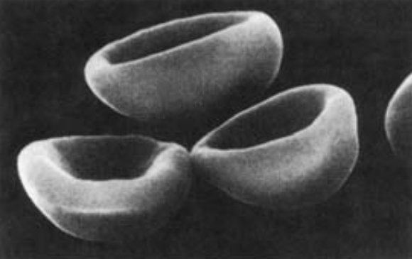
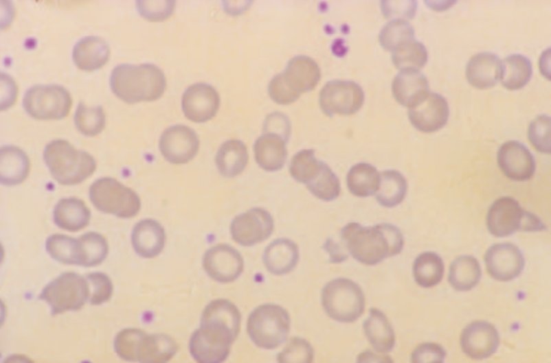
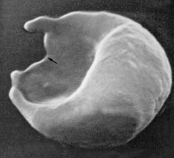
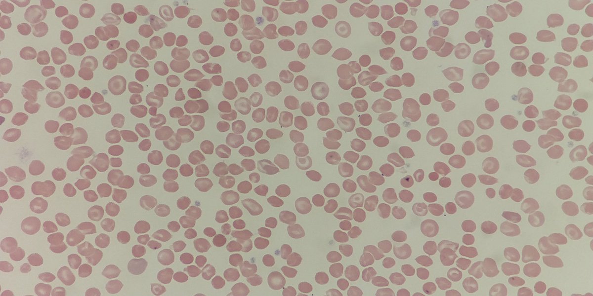
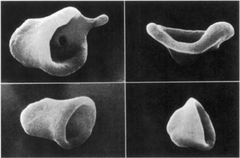
10. Stomatocytes appear having a central linear slit of stoma #underthescope. In wet preps or scanning micrographs, they appear as being cup or bowl shaped. They are noted in vitro (low pH / chlorpromazine use) or in (alcoholic) liver disease amongst others.
#MorphologyMonday
#MorphologyMonday
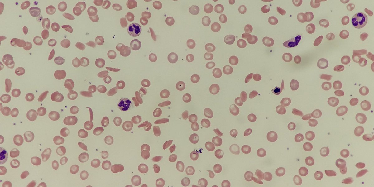
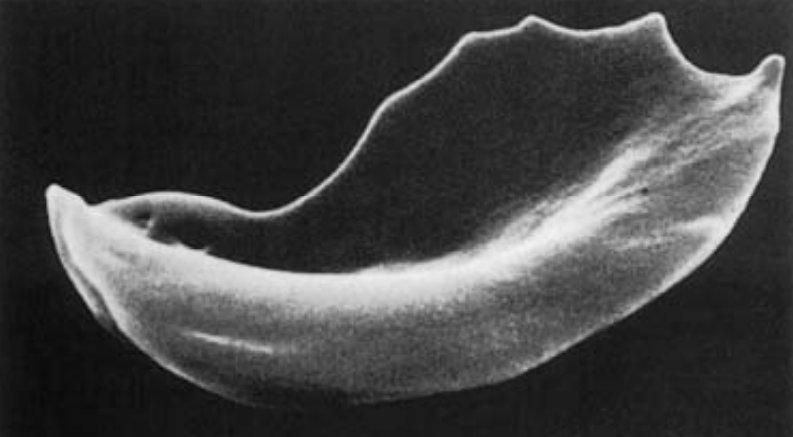
11. #Sickle cells are seen in sickle cell anaemia or sickle cell disease. They are crescent or sickle shaped with pointed ends. #Underthescope they may also appear boat shaped.
#MorphologyMonday
#MorphologyMonday
12. References:
- Blood cells a practical guide, Barbara Bain (+ https://abs.twimg.com/emoji/v2/... draggable="false" alt="📸" title="Camera with flash" aria-label="Emoji: Camera with flash">)
- http://haematologyetc.co.uk/
-">https://haematologyetc.co.uk/">... https://abs.twimg.com/emoji/v2/... draggable="false" alt="📸" title="Camera with flash" aria-label="Emoji: Camera with flash"> : Personal photography
- Blood cells a practical guide, Barbara Bain (+
- http://haematologyetc.co.uk/
-">https://haematologyetc.co.uk/">...

 Read on Twitter
 like appearance. They are thinner than normal cells. In vivo they appear bell shaped but flatten on spreading giving the characteristic target shape. #MorphologyMonday" title="9. Target cells appear #underthescope as having an area of increased staining in the middle giving a https://abs.twimg.com/emoji/v2/... draggable="false" alt="🎯" title="Direct hit" aria-label="Emoji: Direct hit"> like appearance. They are thinner than normal cells. In vivo they appear bell shaped but flatten on spreading giving the characteristic target shape. #MorphologyMonday">
 like appearance. They are thinner than normal cells. In vivo they appear bell shaped but flatten on spreading giving the characteristic target shape. #MorphologyMonday" title="9. Target cells appear #underthescope as having an area of increased staining in the middle giving a https://abs.twimg.com/emoji/v2/... draggable="false" alt="🎯" title="Direct hit" aria-label="Emoji: Direct hit"> like appearance. They are thinner than normal cells. In vivo they appear bell shaped but flatten on spreading giving the characteristic target shape. #MorphologyMonday">
