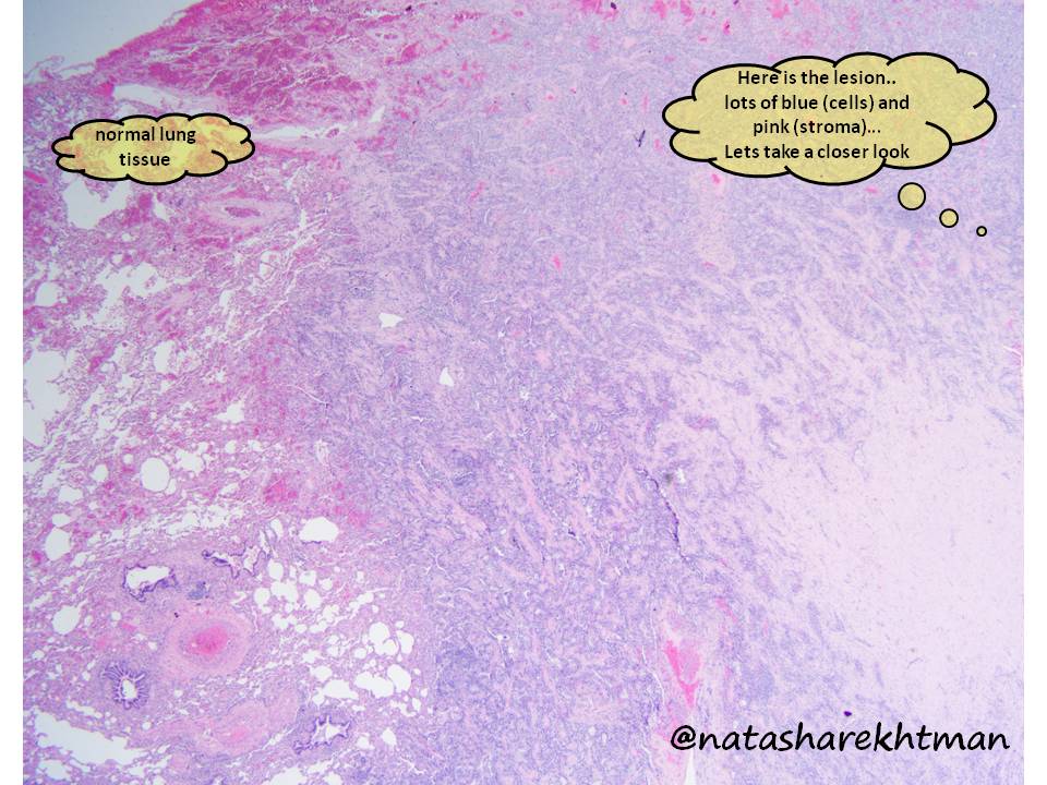
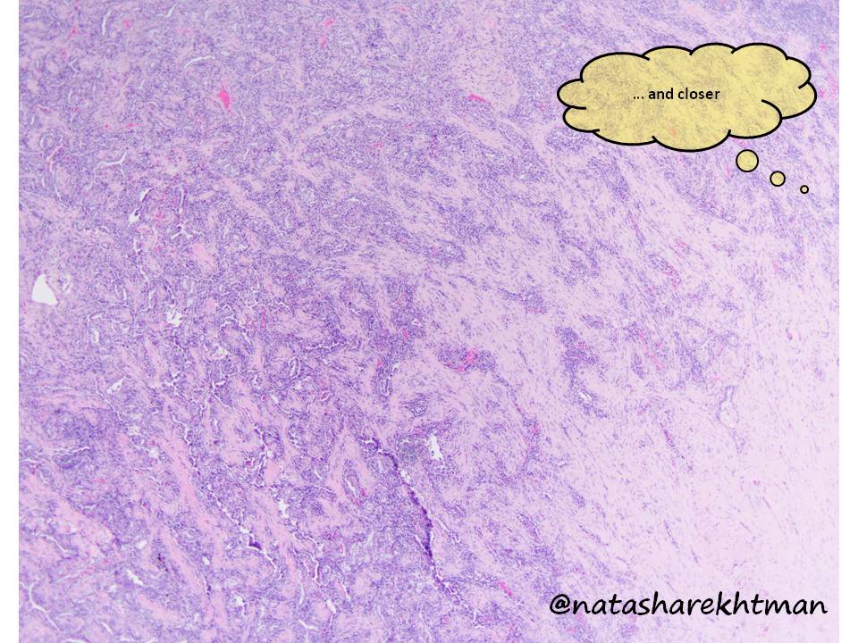
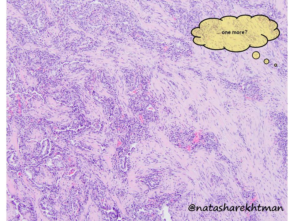
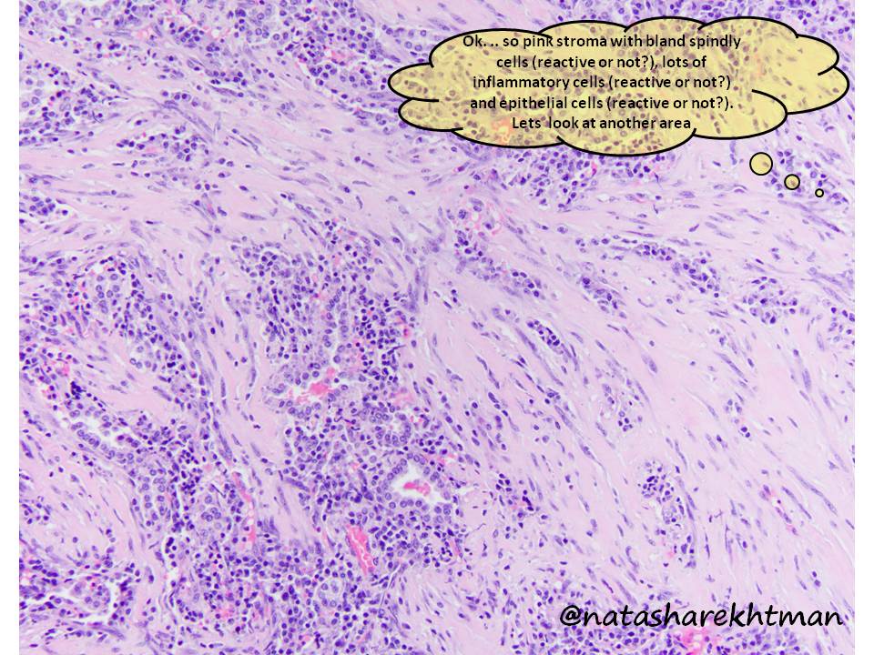
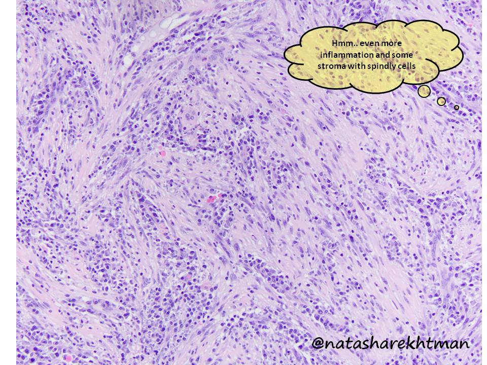
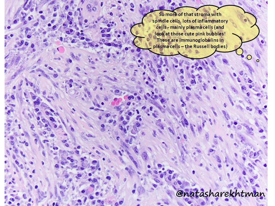
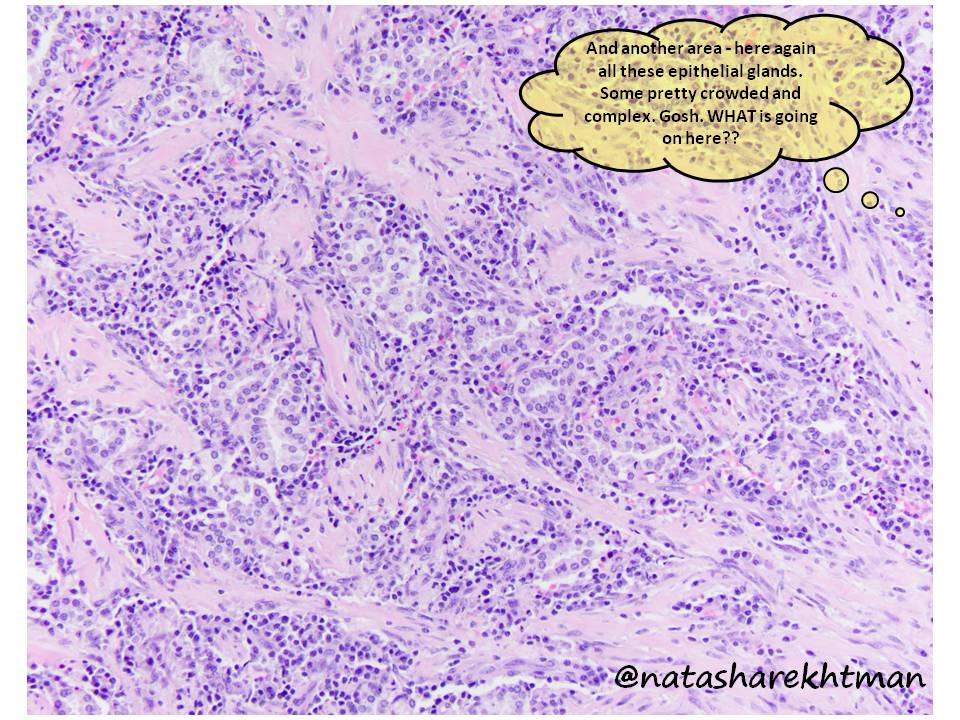
With so much disruption with education, and much need for a distraction… seems like a perfect time for #natpathpuzzler. 3 cm lung nodule in a 40 yo woman. For this one, I included my thoughts as I was first looking at this case (some helpful.. some not). #pulmpath @PulmPathSoc
some more images. This is a case that nicely illustrates a very useful and broad differential. I will post the answer tomorrow... Would love to hear what you would include in your DDx and your best guess (especially from path trainees...)
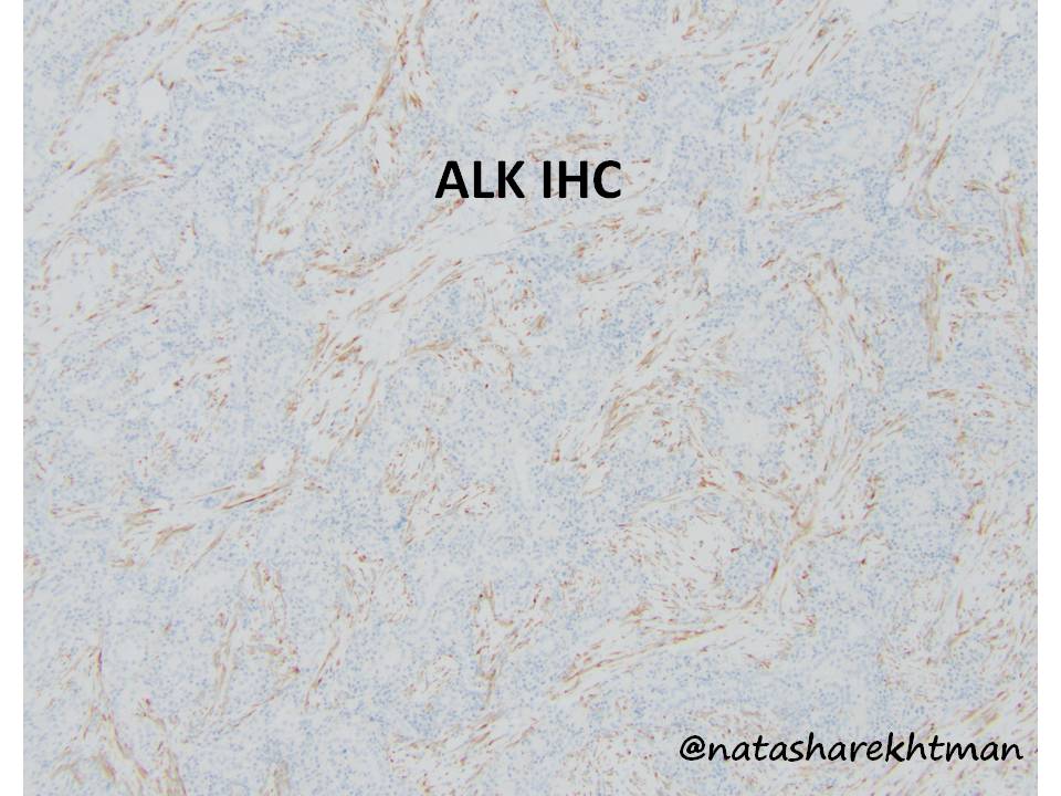
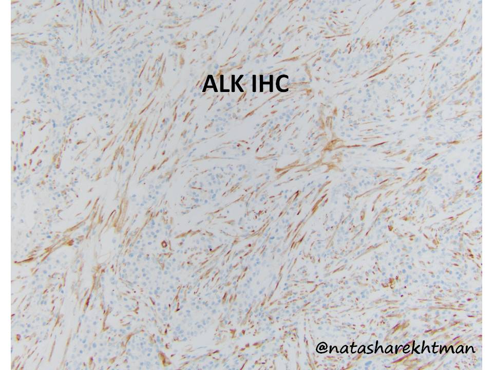
1 Great job everyone! Here is the diagnostic stain: ALK. So this is indeed an inflammatory myofibroblastic tumor (IMT). So lets discuss DDx and a few updates about IMTs.
2 Everyone’s DDx was spot on: IMT #1, IgG4 dz #2, & since many areas were so overrun by lymphoplasmacytic infiltrate – MZL, and lastly (Dx of exclusion… which I never made) – pulmonary hyalinizing granuloma... truly wonder if these might be IMTs before we knew all the markers?)
3 The main point illustrated by this case is that IMTs can be entirely (or mostly) over-run by inflammation & neoplastic spindle cells in some areas can be very inconspicuous. So IMTs should be included in DDx of inflammatory-rich lesions even with very subtle spindle cells.
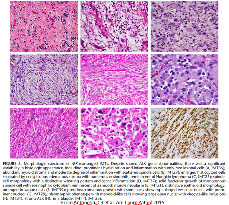
4 Other morphologies of IMTs are well known, and nicely illustrated for various body sites by my great colleague and soft tissue guru Cristina Antonescu. Spectrum ranges from bland, hypocelluar (like our case) to lesions with aggressive features entering in DDx with sarcoma.
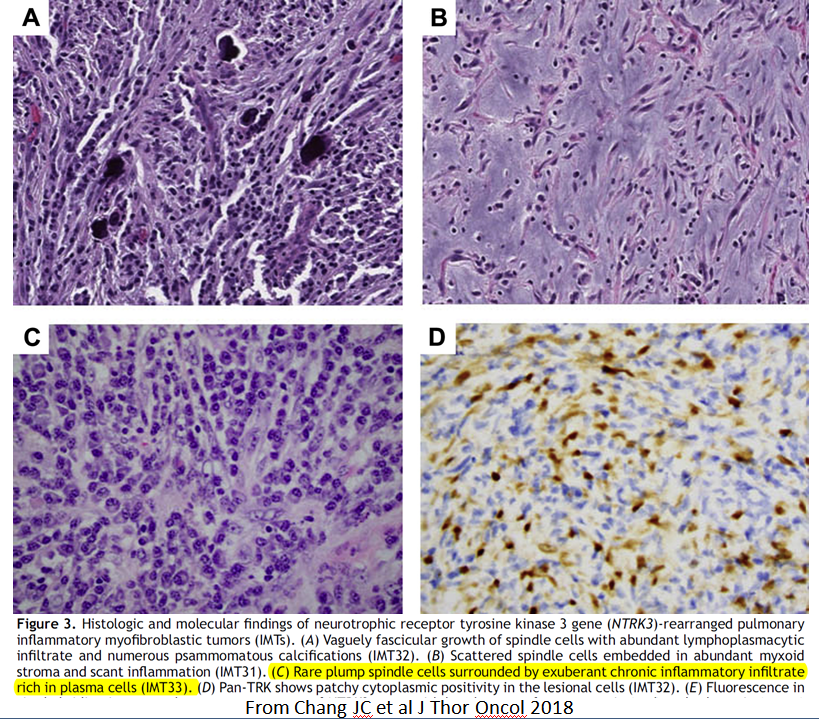
5 In lung, typical IMT morphologies are nicely summarized here. Most classic look is w/ prominent myxoid stroma. But see panel C: as in our case, spindle cells are virtually invisible without IHC! This look of IMT is actually fairly common in lung...now that we know about it!
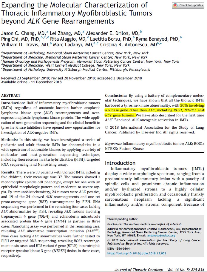
6 What about ALK-negative IMTs? It is well known that ~50% of IMTs have ALK rearrangements (like our case). What about the rest? The answer is fascinating!  https://abs.twimg.com/emoji/v2/... draggable="false" alt="👇" title="Down pointing backhand index" aria-label="Emoji: Down pointing backhand index">
7 Based on a great recent study by my amazing thoracic colleague Jason Chang working with Cristina - with wider gene panel, rearrangements are found in ALL ALK-negative lung IMTs. These include ROS1, RET & NTRK.
8 We have known about some of these based on soft tissue studies https://www.ncbi.nlm.nih.gov/pubmed/25723109 ,">https://www.ncbi.nlm.nih.gov/pubmed/25... but the rate of solved fusions is not as high as in lung. So for possible ALK-neg IMT, always check for these fusions. And if you have ROS1 & pan-Trk IHC – that is a great screening tool.
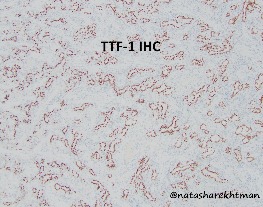
9 Lastly, what about all this epithelial stuff? Here it is highlighted by TTF-1. These are entrapped alveolar structures lined by hyperplastic pneumocytes. When present in the midst of scars, inflammatory lesions, other tumors - they can easily lead you astray. Do not let them!
10 The end - I hope you found this thread educational if you are a trainees or a useful refresher if you are seasoned pathologist. Thanks for your attention!

 Read on Twitter