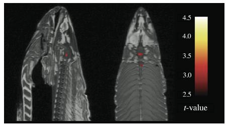
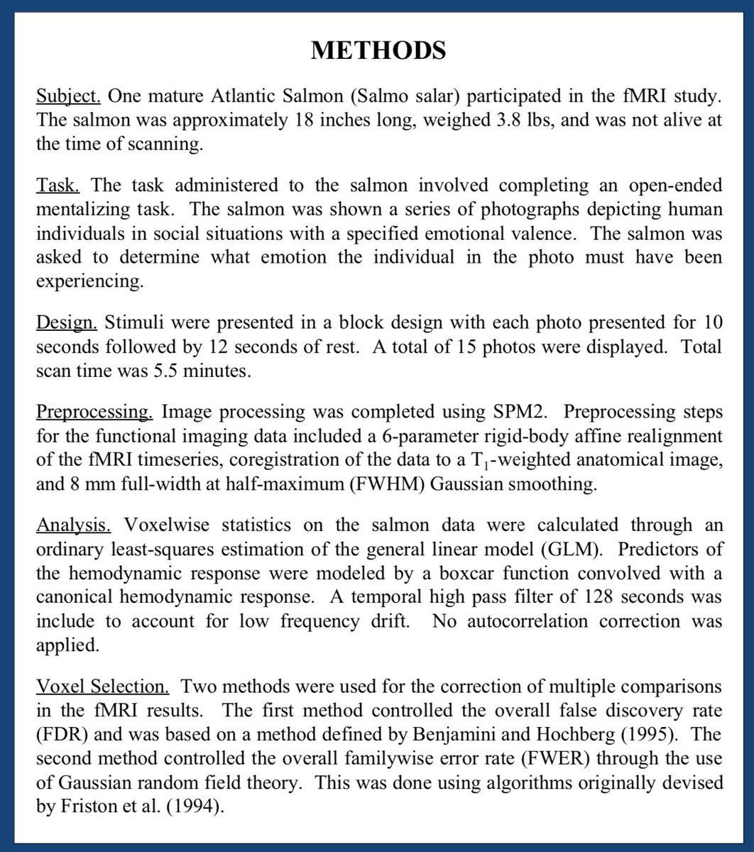
1) In 2008 a salmon (yes the fish) was put through an MRI scanner. The salmon was shown a series of photographs of humans in social situations, and asked to determine the emotion of the person in the photo (yes, really).
2) Running the data through SPM processing showed 3 significant voxels aranged along the midline of the salmon& #39;s brain.
3) Without running through multiple comparisons correction (which isn& #39;t always done) one could conclude that that the salmon was in fact reacting to the photographs.
4) Here& #39;s another fact about that salmon though: it was dead and frozen at the time of the scan.
The experiment was really just a test of the machine. After testing with a pumpkin and a Cornish game hen, they settled on the fish, which had several types of tissue.
The experiment was really just a test of the machine. After testing with a pumpkin and a Cornish game hen, they settled on the fish, which had several types of tissue.
5) As the story goes, the lead author of this study, Dr. Craig Bennett had headed into the store first thing in the morning and proclaimed:
"I need a full length Atlantic Salmon. For science."
"I need a full length Atlantic Salmon. For science."
6) It was not until after a long while, when the another author of the study was running a seminar on how to properly analyze fMRI data, that the significance of the salmon data became a useful example. The salmon study was born.
7) If you do a lot of tests, at least some of them will come out positive, even if they are not real. These are called false positives, and they are something you really want to watch out for.
8) To be clear, the salmon study doesn& #39;t show that fMRI is bogus. It shows the huge importance of correcting your stats.
There are good and bad ways of doing all science.
There are good and bad ways of doing all science.
9) When the poster for this study was presented, between 25-40% of studies on fMRI being published were NOT using corrected comparisons. But by the time these authors won the Ignobel Prize, that number dropped to 10%.
That salmon is a hero.
That salmon is a hero.
10) Reminder: when we see images with areas of the brain in bright colors, that’s not necessarily telling us that one part of the brain is active and the rest isn’t. They’re probability maps. They show the likelihood of activity happening in a given area, not proof of activity.
11) Colorful brain images have powerful persuasive powers. As does most stats.
It& #39;s just generally good advice to understand what you& #39;re looking at.
It& #39;s just generally good advice to understand what you& #39;re looking at.
12) Here& #39;s more info on when they won the IgNobel Prize: https://blogs.scientificamerican.com/scicurious-brain/ignobel-prize-in-neuroscience-the-dead-salmon-study/
And">https://blogs.scientificamerican.com/scicuriou... please also read this interview with two of the authors: it& #39;s brilliant:
https://boingboing.net/2012/10/02/what-a-dead-fish-can-teach-you.html
Craig">https://boingboing.net/2012/10/0... Bennet is also on Twitter: @prefrontal
And">https://blogs.scientificamerican.com/scicuriou... please also read this interview with two of the authors: it& #39;s brilliant:
https://boingboing.net/2012/10/02/what-a-dead-fish-can-teach-you.html
Craig">https://boingboing.net/2012/10/0... Bennet is also on Twitter: @prefrontal
13) Moral of the story: don& #39;t go fishing unless you& #39; re prepared to not catch any fish.
http://prefrontal.org/files/posters/Bennett-Salmon-2009.pdf">https://prefrontal.org/files/pos...
http://prefrontal.org/files/posters/Bennett-Salmon-2009.pdf">https://prefrontal.org/files/pos...
14) If you enjoyed this thread you may also appreciate my thread on the replication crisis: https://twitter.com/axbom/status/956829916548550656?s=21">https://twitter.com/axbom/sta...
15) https://twitter.com/axbom/status/1203595984246325248?s=21">https://twitter.com/axbom/sta...
16) To everyone saying that scanning live salmon wouldn& #39;t work anyway, it has been done :) https://www.discovermagazine.com/mind/fmri-scanning-salmon-seriously">https://www.discovermagazine.com/mind/fmri...
17) To be even more clear: fMRI does not measure activity in the brain. You& #39;d need electrodes implanted in the brain to do that. It measures magnetic disruption in the brain, based on oxygenated and deoxygenated blood having different magnetic properties. Hence false positives.
18) "There& #39;s all kinds of noise that gets entered into the signal. It& #39;ll pick up your own heart beating. We once had a lightbulb going bad in the scanner suite that was introducing specific signal in our data set. You have to get enough data [...] to separate signal from noise."
19) "More than a couple papers have been sesationalistic. There have been comparisons of Republican and Democratic brains. That& #39;s ridiculous and it& #39;s a misuse of fMRI."
Do read the interview with the authors if you didn& #39;t already. https://boingboing.net/2012/10/02/what-a-dead-fish-can-teach-you.html/amp?__twitter_impression=true">https://boingboing.net/2012/10/0...
Do read the interview with the authors if you didn& #39;t already. https://boingboing.net/2012/10/02/what-a-dead-fish-can-teach-you.html/amp?__twitter_impression=true">https://boingboing.net/2012/10/0...

 Read on Twitter


