Membranous-like Glomerulopathy with Masked IgG kappa Deposits (MGMID) is an immune complex-mediated kidney disease, usually affecting young women. This work is by @renalpathdoc and @NephroRock, who recently identified a useful biomarker for MGMID, serum amyloid P (SAP).
This tweetorial includes data from the following papers, all from @arkanalabs :
https://www.ncbi.nlm.nih.gov/pubmed/24429395
https://www.ncbi.nlm.nih.gov/pubmed/24... href=" https://www.ncbi.nlm.nih.gov/pubmed/29142932
https://www.ncbi.nlm.nih.gov/pubmed/29... href=" https://www.kidney-international.org/article/S0085-2538(19)31119-6/pdf">https://www.kidney-international.org/article/S...
https://www.ncbi.nlm.nih.gov/pubmed/24429395
MGMID is a very rare renal disease, making up only 0.1% of kidney biopsies (41/42,711 biopsies in a 5 year period).
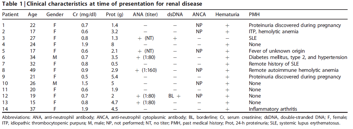
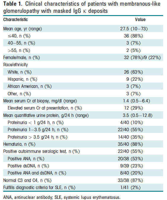
Some patients with MGMID have a positive ANA or vague autoimmune manifestations, but most do not have a clinical diagnosis of SLE. They typically undergo kidney biopsy for proteinuria, which can be nephrotic range.
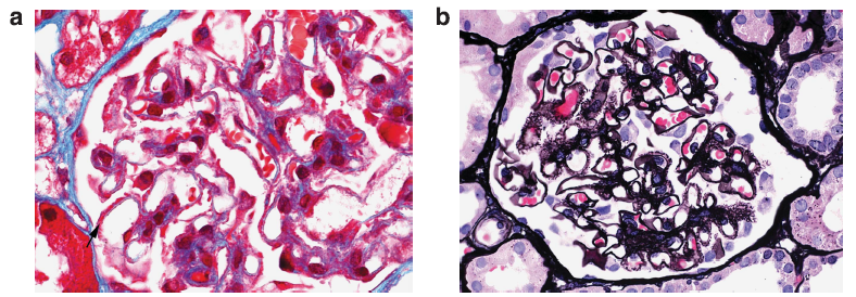
On light microscopy, the glomerular capillary loops appear prominent and there are fuscinophilic deposits on trichrome stain (a) and may be the formation of spikes and holes on silver stains (b), as also seen in membranous glomerulopathy (it’s membranous-like).
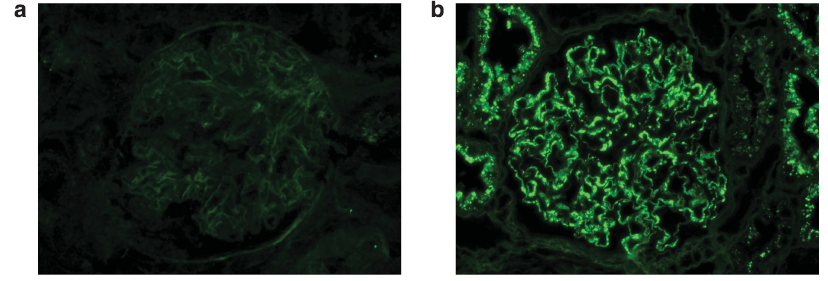
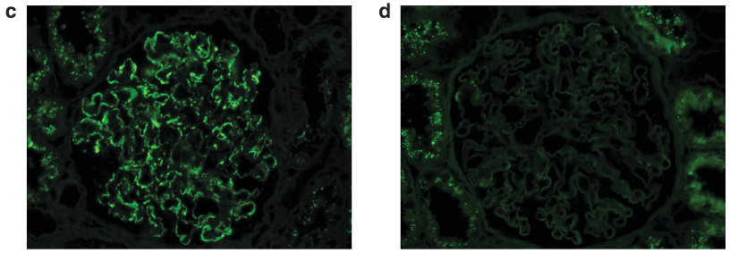
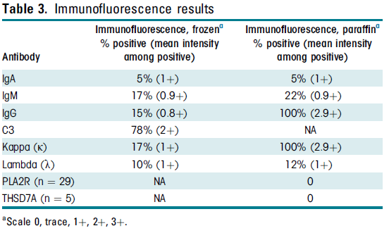
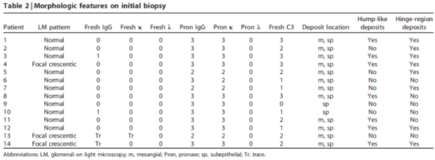
On immunofluorescence microscopy, IgG is negative in a majority of cases (a). However, pronase digestion on FFPE tissue will lead to “unmasking” of immune deposits, which will be positive along the glomerular capillary loops and frequently also within the mesangium (b).
There is unmasked kappa (c), but not lambda (d), light chains in the same distribution as IgG on paraffin immunofluorescence.
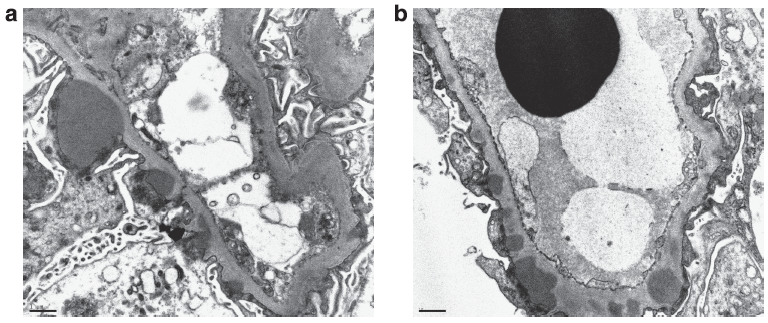
Electron microscopy often shows subepithelial and intramembranous deposits, and frequently also shows mesangial immune deposits.
You should consider a diagnosis of MGMID when there is only C3 staining on routine immunofluorescence, but a membranous-like pattern by electron microscopy. Performing paraffin IF will unmask immune deposits.
Some patients will not have masked IgG kappa and will have deposits seen on routine immunofluorescence. In these cases, IgG subclasses can be performed, and the deposits will be IgG1 restricted.
MGMID can be distinguished from PGMID by the lack of endocapillary proliferation and subendothelial deposits. Unlike monoclonal membranous glomerulopathy, MGMID typically has masked IgG deposits.
A larger cohort of 41 patients showed female predominance (78%) and a clinical presentation with proteinuria, microscopic hematuria, and often a positive ANA without clinical features of lupus.
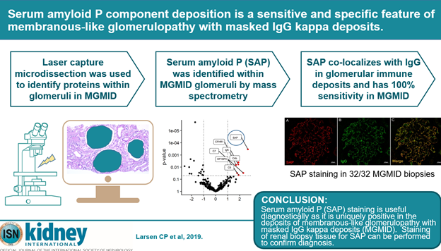
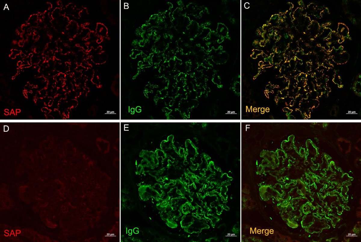
Mass spectrometry and immunohistochemistry revealed that SAP is uniquely present in glomerular deposits from MGMID.
SAP was expressed in a granular capillary loop (and sometimes mesangial) pattern within glomeruli and showed co-localization with IgG within glomerular deposits (c). PLA2R positive MN is shown in the bottom panels as a negative control, which shows no staining within glomeruli.
SAP staining of multiple glomerular diseases, including C3G, DDD, fibrillary, IgAN, PLA2R or THSD7A positive MN, infection-associated GN, cryo-GN, membranous or proliferative lupus nephritis, and PGMID were all neg & were positive in all cases of MGMID, showing high specificity.
The reason for the light chain restriction in this entity is currently unknown but this is not thought to be a monoclonal gammopathy of renal significance (MGRS) lesion.
SAP staining by paraffin immunofluorescence of kidney biopsies is a useful diagnostic tool for MGMID, which is both sensitive and specific. To learn more about SAP in MGMID, check out https://www.kidney-international.org/article/S0085-2538(19)31119-6/pdf">https://www.kidney-international.org/article/S...

 Read on Twitter