Trainees! #MedStudents, #Residents, #Fellows - let& #39;s talk about the #neuromodulation kit and its contents. This is the stuff no one will show you, but on your first case you will be expected to have a general understanding. Let& #39;s get started (follow along):
There will be a 14 gauge introducer needle with an inserted stylet. Using the next lower ipsilateral pedicle as your starting point, you simply get loss of resistance at your desired level.
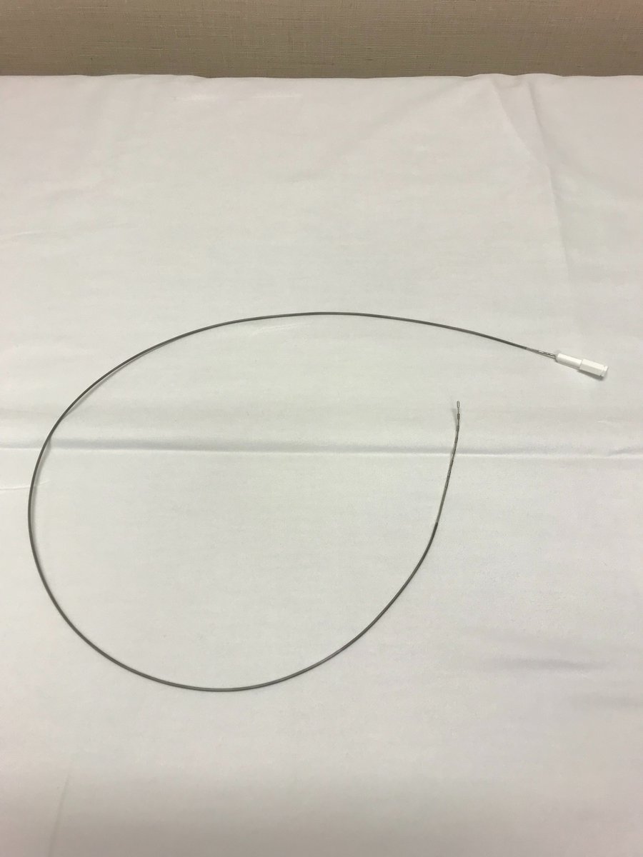
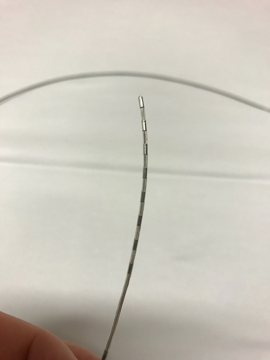
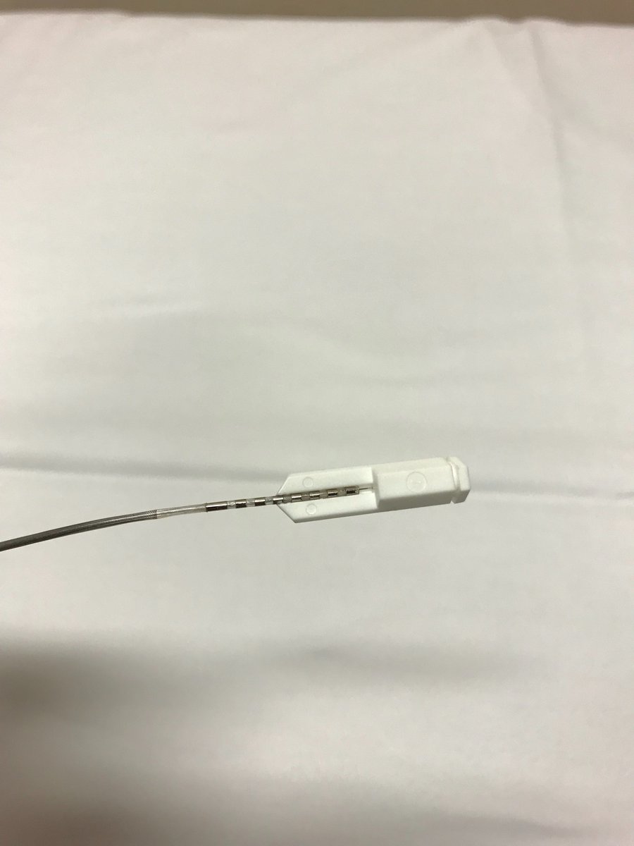
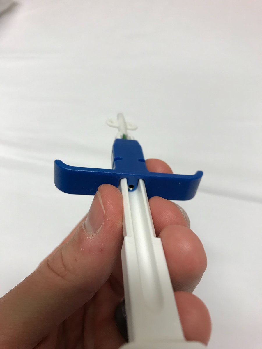
Next, you thread a lead through the introducer needle into the epidural space (picture 1). The lead has, in this case, eight electrodes at its tip (picture 2). The opposite end has contact points for these electrodes (picture 3).
The picture 3 end is attached to an internal stylet for the lead. This helps with "driving" the lead into the appropriate position. This typically requires some manipulation, which is accomplished with rotation of the internal stylet. A slight bend in the stylet helps.
After getting the leads into appropriate position using fluoroscopy and lead manipulation, the lead internal stylet is removed. If this were a trial, the leads would be attached to the skin, hooked to a battery, and the case is over.
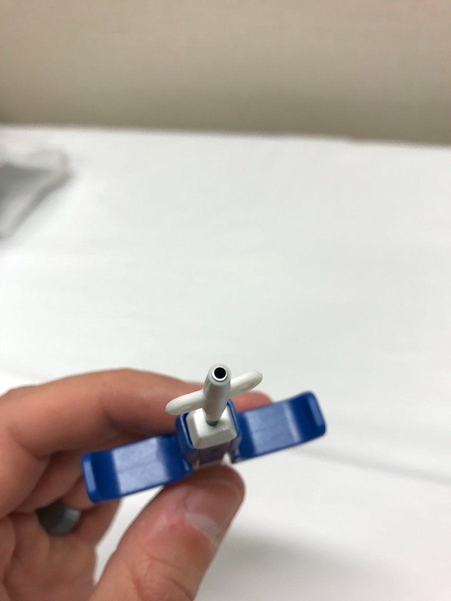
If its an implant though, there are a few more steps. The leads must be secured to the underlying fascia. There are many systems and methods to accomplish this. One design is the "bi-winged anchor". This system by @Medtronic is particularly easy to use.
The lead is slid into the end of the device (picture 1). Upon threading the lead into this device, it will emerge from the other end (picture 2). The tool is slid down to the fascia and deployed. The white anchor is left behind and the rest of the device is removed.
The lead is strongly adhered to the anchor at this point and the surgeon simply sutures the anchor using the "wings" to the fascia.
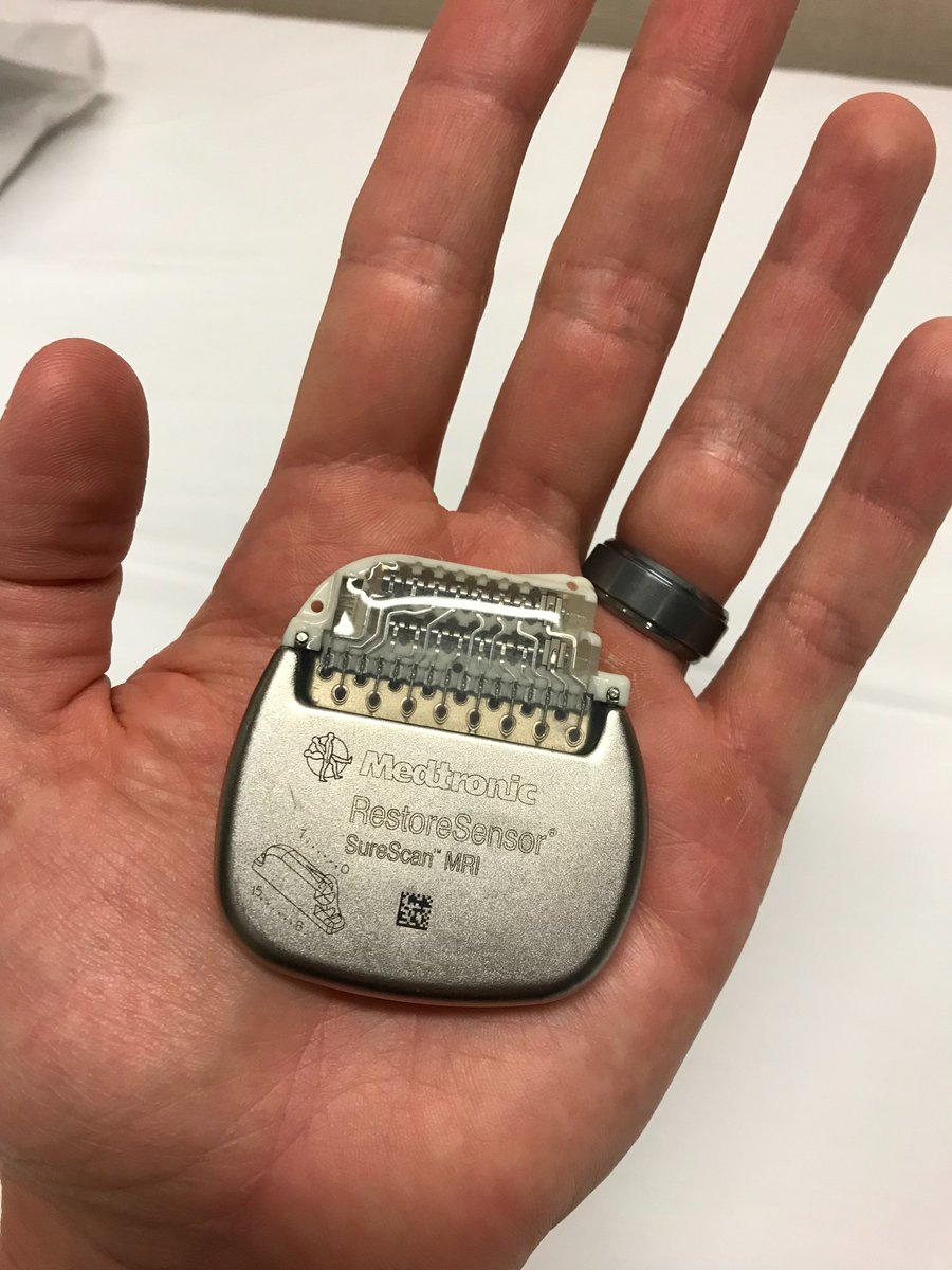
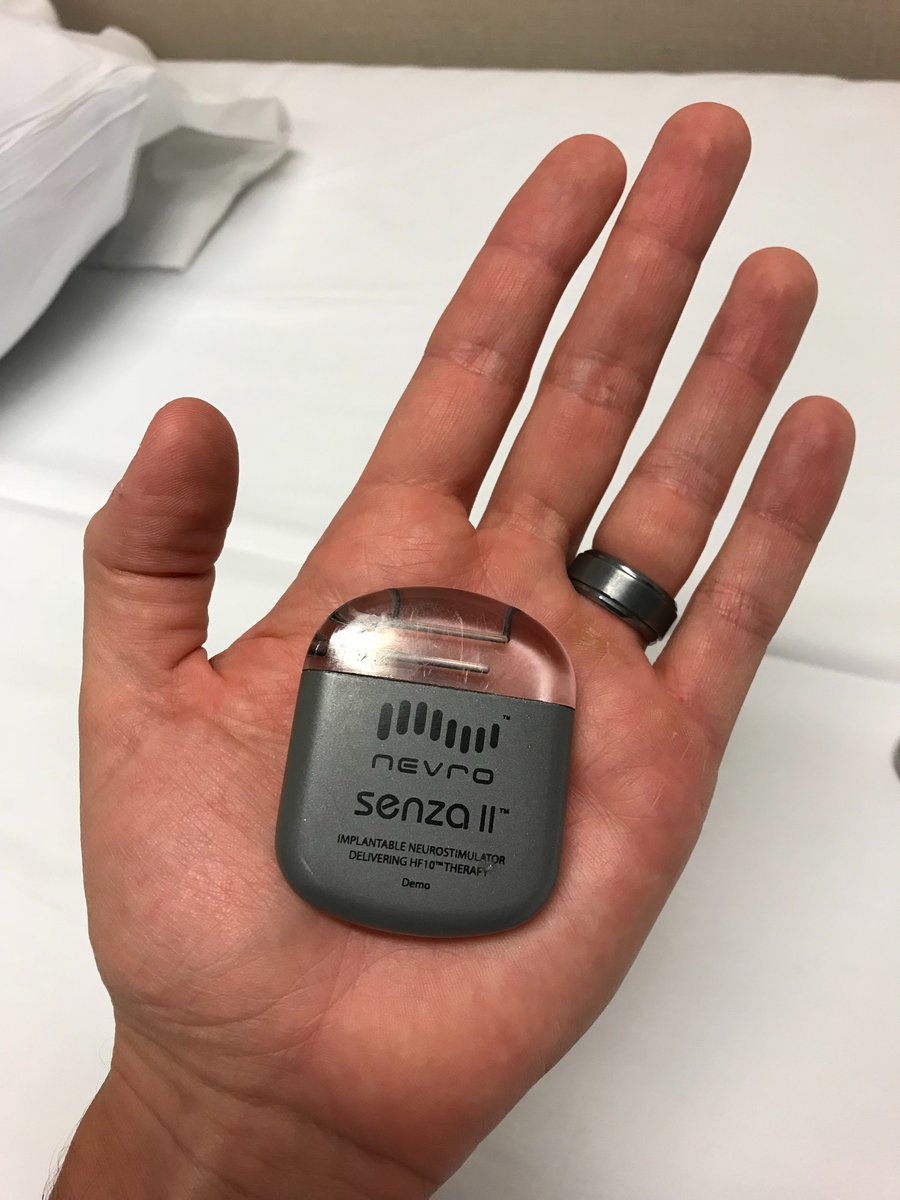
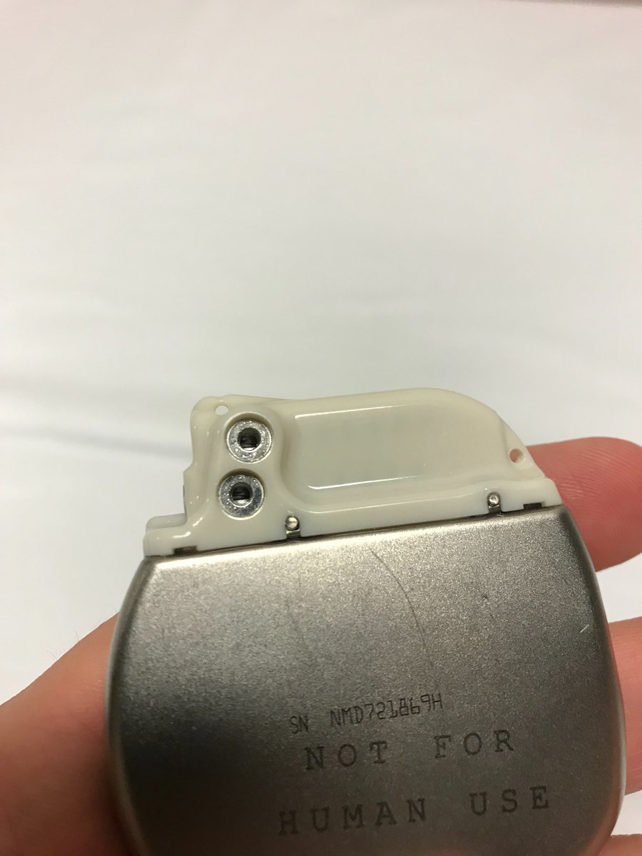
The leads are tunneled beneath the skin to the site of battery location, i.e. "the pocket". The battery is typically referred to as the #IPG - implantable pulse generator. Each company has a unique size, shape, and characteristics.
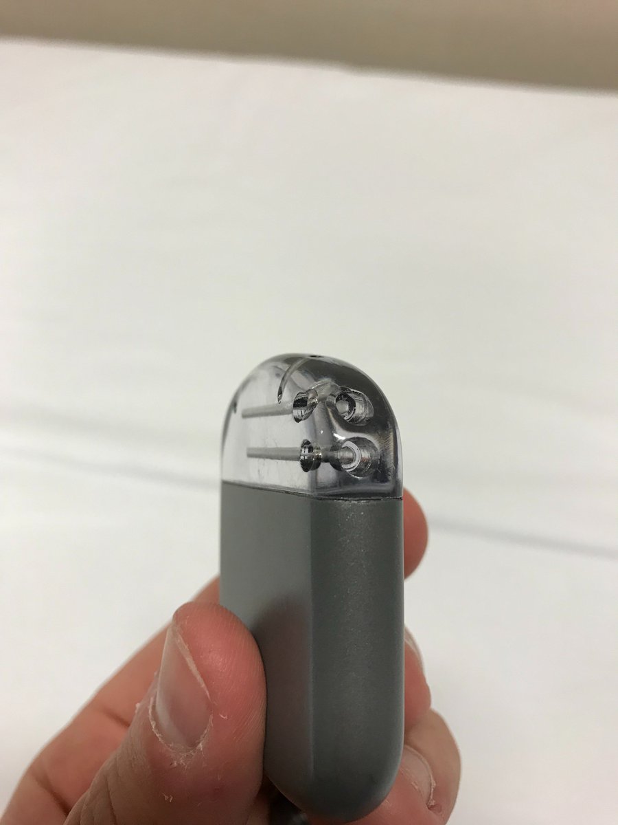
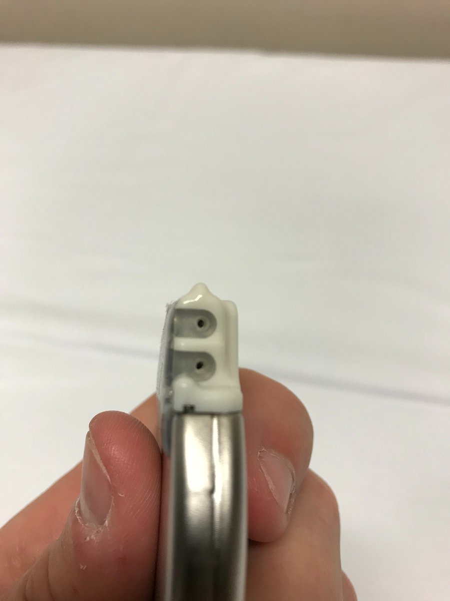
The contact point end of the leads are placed inside of the IPG. The leads are slid into position (pictures 1,2), and secured with a small screw and screwdriver (picture 3). At this point, the electrodes in the epidural space have been placed in continuity with the IPG.
The leads are coiled into loops, and along with the IPG, are placed into the pocket. The skin incision over the spine and the pocket are irrigated well, hemostasis is ensured, and the incisions are closed appropriately. The incisions are covered with a dressing.
This completes the case. The patient is brought to the PACU and after they are safe to go home, they are discharged. You will discharge them with abx and possibly pain meds.
I hope this helps! If you have any questions, just reply to this thread. #meded #traineesasteachers
I hope this helps! If you have any questions, just reply to this thread. #meded #traineesasteachers

 Read on Twitter